Female Pelvic Floor
On this page
Related
Further Reading
Functional UrologyFurther Reading · Voiding DysfunctionAnatomyFurther Reading · BasicsReconstructionFurther Reading · Reconstructive- In uterosacral ligament suspension, the ureter lies lateral to the ligament's anterior portion (closest at the cervix), and the S1 and S2–S4 nerve trunks cross dorsally, risking sacral nerve entrapment.
- DeLancey level I (apical) support, the cardinal and uterosacral ligaments, suspends uterus, cervix and upper vagina to sacrum and pelvic side wall; hysterectomy endangers it, and loss causes apical (vault) prolapse, often with anterior prolapse.
- Loss of DeLancey level II (mid-vagina to arcus tendineus fascia pelvis) causes cystocele, while loss of level III (perineal membrane, levator ani, perineal body) is linked to distal posterior-wall and perineal defects.
- POP-Q references all measurements to the hymen, not the introitus, because the hymen can be located more precisely, and staging is done in dorsal lithotomy with the patient straining.
- POP-Q stage follows the leading edge, the most positive of Aa, Ba, Ap, Bp and C: Aa -2, Ba 0, C -5, Ap +1, Bp +1 gives leading edge +1, stage 2.
- POP-Q stages by leading edge: 1, more than 1 cm proximal to the hymen; 2, within 1 cm of it; 3, more than 1 cm beyond it without total vaginal eversion; 4, complete eversion.
Support Anatomy — DeLancey's 3 Levels
| Level | Supports | Structures | Damage → |
|---|---|---|---|
| I (upper) | Upper vagina (+ uterus) | Cardinal / uterosacral ligaments — connective tissue suspending the apex to the sacrum and lateral pelvic side wall | Vaginal apex prolapse (uterine / vault) |
| II (middle) | Mid-vagina | Fascial arcus: pubocervical fascia anteriorly · rectovaginal fascia posteriorly · levator ani (through the arcus tendineus fasciae pelvis) | Cystocele or rectocele |
| III (lower) | Lower vagina | Perineal membrane · urogenital diaphragm — fusion of the anterior vagina with the urethral support muscles, and of the posterior vagina with the perineal body | Anterior → urethrocele · posterior → deficient perineal body |
- Level I suspends the uterus, cervix and upper vagina
- Its ligaments are at risk during hysterectomy → post-hysterectomy vault prolapse
- Loss of apical support lets the anterior wall descend too → apical + anterior prolapse often occur together
- Level III also fuses the lower vagina with the levator ani; its loss → distal posterior-wall and perineal defects
- ATFP runs pubic bone → ischial spine; separation from it = paravaginal defect
- Urogenital hiatus held closed by tonic levator tone
- Fascia carries almost no load until the muscle fails, then ligaments take everything and fail over time
- Levator avulsion in 23% of symptomatic women → higher stage, three-compartment POP
- Visible levator defects on MRI in 20% of primiparas, none in nulliparas
- Levator innervation S3–S5; pudendal supplies urethral/anal sphincters
- Ehlers-Danlos and Marfan → increased POP risk (altered collagen I:III)
Integral theory — the 3 supports of the proximal/midurethra
- Pubourethral ligaments
- Suburethral vaginal hammock
- Pubococcygeus muscle
- ISD is the primary cause of female SUI; hypermobility is secondary — all women with SUI have some ISD, not all have hypermobility
- Exam answer: ISD is likely present in the majority of women with SUI
- Hypermobility is secondary because many women have it and stay continent
- Hypermobility is a symptom of damaged support, not a cause of SUI
- In continent women MUCP occurs at the midurethra
Pelvic Organ Prolapse — Basics
| Compartment | Organ | POP-Q points |
|---|---|---|
| Anterior | Bladder (cystocele), urethra | Aa, Ba |
| Apical | Uterus, cervix, vault, bowel (enterocele) | C, D |
| Posterior | Rectum (rectocele), small bowel | Ap, Bp |
- Frequency: anterior > posterior > apex (least common) — but apex is under-diagnosed, not rare
- Enterocele = true hernia of intestine into the vaginal wall
- Procidentia → bilateral ureteric obstruction — relieved by reducing the prolapse
- Apical descent (C ≥ −3) present in 42% / 85% / 100% of stage 2 / 3 / 4 anterior POP → suspect it before every cystocele repair
- Prevalence 3–6% by symptoms, up to 50% anatomically; lifetime risk of POP surgery 12.6%
- Asymptomatic stage 1–2 is normal — symptoms rarely appear until the prolapse reaches the hymen
- Natural history slow: at 16 months 19% progress · 78% no change · 3% regress
- Progression risk higher if age >60, Ba ≥4 cm, or genital hiatus ≥5 cm
Risk factors
- Well-established (3): age · obesity · parity
- Parity carries the strongest association with the development of POP
- Adjusted RR 8.4 after 2 deliveries, 10.85 after 4
- Each extra delivery up to 5 births adds 10–20%
- Forceps OR 5.4 vs vaginal delivery OR 2.9 for POP surgery
- Cesarean section appears protective against prolapse
- Same three for pelvic floor disorders generally
- Parity is the one most specific to sphincter damage → prolonged labor · third-degree tears · high birth weight · forceps delivery
- Parity carries the strongest association with the development of POP
- Less established (10):
- Smoking
- Chronic constipation
- Menopause
- Hysterectomy/pelvic surgery
- Hysterectomy done for POP strongly predicts repeat pelvic floor surgery
- Heavier vaginally delivered fetus
- Genetic
- Race
- Hispanic women have the highest prevalence, Black women the lowest; more common in Caucasian and Hispanic than African-American women
- Heavy lifting
- Intense physical activity
- Chronic pulmonary disease
POP-Q
| Point | Definition | Range |
|---|---|---|
| Aa | Anterior wall 3 cm proximal to meatus (≈ bladder neck) | −3 to +3 |
| Ba | Most distal point of anterior wall prolapse | −3 to +tvl |
| Ap | Posterior wall 3 cm proximal to meatus | −3 to +3 |
| Bp | Most distal point of posterior wall prolapse | −3 to +tvl |
| C | Cervix (or vaginal cuff) | −10 to +10 or more |
| D | Posterior fornix (only if cervix present) | −10 to +10 or more — not recorded after hysterectomy |
| gh | Mid-meatus → posterior midline hymen | — |
| pb | Posterior gh margin → middle of anus | — |
| tvl | Total vaginal length, fully reduced | — |
- Hymen = the reference point (more precisely identified than the introitus); negative = above, positive = below
- All points measured while straining
- Staging exam done in dorsal lithotomy
- C vs D distinguishes cervical elongation from true uterine prolapse
- Wide C–D gap (e.g. C 0, D −5) = elongated cervix → hysteropexy / sacrouteropexy leave it unaddressed
- Ba and Bp can reach +5 or +6 in extreme prolapse — the
+tvlceiling is not a small number
| Stage | Leading edge | Formal criterion |
|---|---|---|
| 0 | No prolapse | Aa, Ap, Ba, Bp all at −3, and C or D ≤ −(tvl − 2) |
| 1 | >1 cm above the hymen | Stage 0 criteria not met and leading edge < −1 cm |
| 2 | Within 1 cm either side of the hymen | Leading edge ≥ −1 cm but ≤ +1 cm |
| 3 | >1 cm below the hymen, not totally everted | Leading edge > +1 cm but < +(tvl − 2) |
| 4 | Complete eversion | Leading edge ≥ +(tvl − 2) |
- Stage follows the leading edge = the most positive of Aa, Ba, Ap, Bp, C
- e.g. Aa −2, Ba 0, C −5, Ap +1, Bp +1 → leading edge +1 → stage 2
- Do NOT measure immediately after removing a vaginal pessary — it underestimates the true degree of prolapse
POP — Evaluation
- Vaginal bulge is the ONLY symptom strongly associated with prolapse at or below the hymen
- Everything else correlates weakly with site and severity
- Examine in lithotomy AND standing; if symptoms and stage disagree, re-examine standing or later in the day
- Signs of estrogen deficiency (3): urethral caruncle · urethral prolapse · labial adhesions
- Anal sphincter tone = S2–4
- No routine imaging
- But dynamic MRI adds real information pre-operatively when the pelvic examination is difficult or inconclusive
- Evaluate defecatory dysfunction before operating
POP and incontinence
- >40% of women with SUI have a significant cystocele
- POP surgery improves storage symptoms: frequency 36.6 → 14.6%, urgency 36.8 → 12.9%, UUI 21.2 → 6.1% (de novo 5–6%)
- Anti-incontinence surgery without correcting POP can worsen POP
- Advanced untreated POP → retention (urethral compression) and rarely renal failure (ureteric compression)
Occult SUI — the pre-op question
- SUI appearing only after prolapse reduction, previously masked by high-stage anterior POP
- Exam answer: marked prolapse → risk of de novo or worsening SUI after repair
- Detection 4.4% unreduced → 22% with pessary reduction
- 29.2% of symptom-free women with POP leak on at least one of five stress tests
- Do a POP reduction test in every continent woman before POP surgery (EAU Strong)
- If significant POP, run UDS with AND without a pessary
- Additional evaluation indicated if POP-Q ≥3 and SUI not demonstrated on reduction
- A vaginal pack works as well as a pessary for reducing the prolapse during the study
- Run a filling study (CMG) for detrusor overactivity and measure VLPP during the reduced study — not just a stress test
- If manual reduction during UDS produces no leak, a pessary adds nothing — do not repeat it
- Overfilling the bladder beyond capacity adds nothing either; no reduction method is standardized
- Remove any urethral catheter and reassess — an indwelling catheter can itself mask SUI
- Order:
- Subjective SUI + high-grade (stage 3) POP, no leak on UDS → repeat stress testing with the prolapse reduced first
- Still dry → remove the urethral catheter and repeat stress maneuvers with the prolapse still reduced — the catheter masks SUI by obstruction (AUA/SUFU)
- Order:
POP — Management
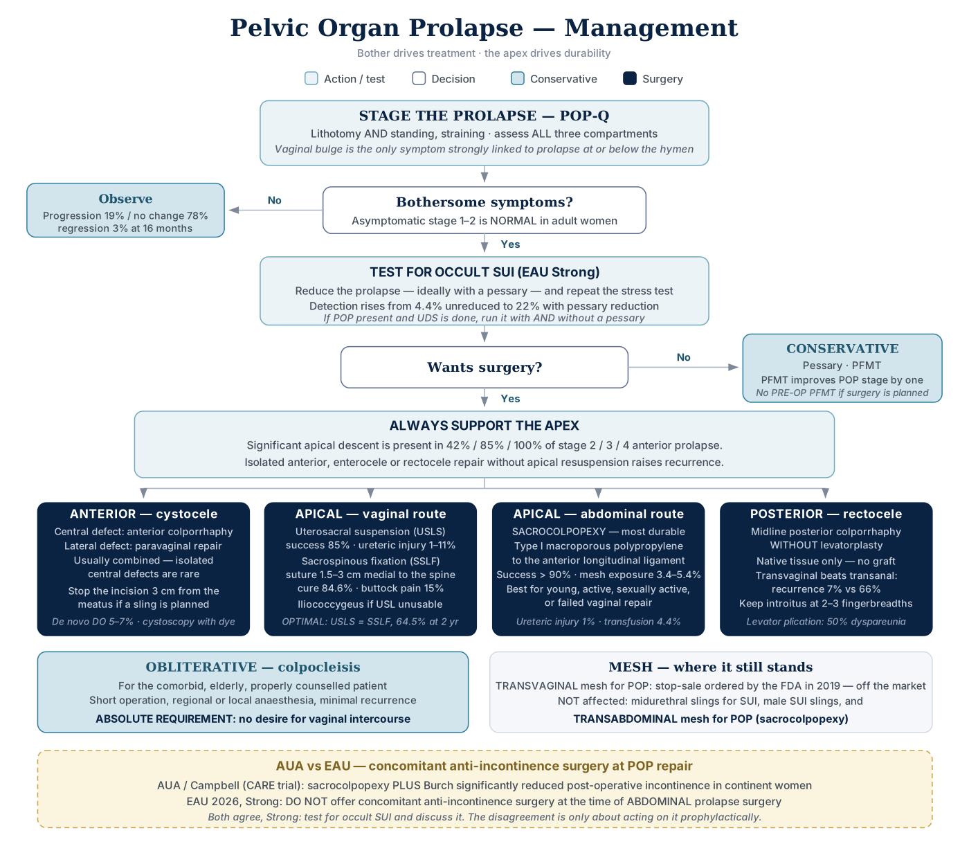
Click the algorithm to open it full size.
Conservative
- Options: observation · pessary · PFMT
- PFMT improves POP symptoms and/or POP stage by one stage in stage I–III
- Do NOT give pre-operative PFMT if a pessary or surgery is already indicated
- 10 RCTs show no benefit
- OPTIMAL found peri-operative PFMT did not improve outcome
- Urgency incontinence refractory to antimuscarinics + asymptomatic anterior wall prolapse to the hymen → pessary trial first
- Shows whether correcting the prolapse improves the symptoms
- Improves → continue the pessary or proceed to cystocele repair
- No help → sacral neuromodulation or other refractory OAB options
Three surgical families
| Type | Principle | Best for |
|---|---|---|
| Restorative | Repair discrete defects with native tissue | Reasonable tissue, discrete defects |
| Compensatory | Reinforce with a graft | Weak tissue, failed prior repair |
| Obliterative | Close the vagina | Comorbid, elderly, not sexually active |
- The first operation is the best chance — recurrence rises with each attempt
- Vaginal: fewer complications, shorter stay ⟷ abdominal: more durable
- Success = symptom resolution, not anatomy
- Patients perceive catheters, constipation and urgency as complications — counsel explicitly
ALWAYS SUPPORT THE APEX. Isolated anterior, enterocele or rectocele repair without apical resuspension increases recurrence.
Anterior — cystocele
- Central defect → anterior colporrhaphy · lateral defect → paravaginal repair
- Usually combined; isolated central defects are rare
- Post-hysterectomy stage 3 anterior prolapse with apical descent → vault suspension + anterior repair
- Anterior repair alone has unacceptably high failure rates
- Midline incision to 1.5 cm from the meatus
- But stop 3 cm from the meatus if a sling is planned (sling needs its own midurethral incision)
- Plicate 2-0 delayed absorbable, tension-free, stay shallow to avoid the bladder → cystoscopy with dye
- Complications:
- De novo DO 5–7% (but pre-existing DO resolves in up to 63%)
- Retention
- Bleeding >350 mL
- Bladder/ureteric injury
- VVF
- Dyspareunia
- Bleeding usually = wrong plane — dissect on the white shiny surface of the pubocervical fascia
- No dye efflux → remove the plication sutures
Apical — vaginal route
| Procedure | Key technical point | Results | Signature complication |
|---|---|---|---|
| Uterosacral suspension (USLS) | 3 permanent sutures per USL, proximal and medial to the ischial spine; cystoscopy before cutting sutures | Success 85% (48–96%); reoperation 5.8%; by compartment ant 81.2% / apical 98.3% / post 87.4% | Ureteric kinking or injury 1–11% |
| Sacrospinous fixation (SSLF) | Suture 1.5–3.0 cm medial to the ischial spine — too close risks Alcock's canal, too medial risks failure and sacral nerves | Cure 84.6%; apical recurrence 5.3%; anterior recurrence 18.3% | Ipsilateral buttock pain 15%, resolves 2–3 months |
| Iliococcygeus suspension | Fixation 1 cm distal to the ischial spine; bilateral | Cure 53–96% | Buttock pain |
- The ischial spine is the most important landmark in apical prolapse surgery
- It orients every vaginal apical suspension
- USLS anatomy:
- The ureter lies lateral to the anterior portion of the USL, closest at the cervix
- Highest reported ureteric injury risk of the prolapse procedures
- Exam answer: 0–11% · vs transvaginal hysterectomy 0.2 per 1000 · uncommon with anterior colporrhaphy
- Highest reported ureteric injury risk of the prolapse procedures
- The S1 and S2–S4 nerve trunks cross dorsally → risk of sacral nerve entrapment
- Exam answer: pass the sutures at the level of the ischial spine (the USL lies posterior and medial to it) — placement beyond a mean 4.6 cm from the spine toward the sacrum → S3 nerve injury
- The ureter lies lateral to the anterior portion of the USL, closest at the cervix
- SSLF anatomy: too close to the spine → pudendal entrapment
- The pudendal nerve and vessels lie directly behind the ischial spine
- Also keep clear of the area above and just behind the ligament (inferior gluteal vessels, hypogastric venous plexus)
- The sacral plexus lies immediately behind the ligament as it leaves through the greater sciatic foramen → posterior and lower leg pain after SSLF = sacral plexus injury (the pudendal nerve can also be injured)
- Significant bleeding during SSLF → most likely from vessels of the posterior division of the internal iliac artery
- SSLF deflects the vagina posteriorly (and right if unilateral) → anterior compartment recurrence
- OPTIMAL trial: USLS vs SSLF — no difference, 2-year success 64.5%
- Absorbable suture is non-inferior to permanent for apical recurrence
- Concomitant anterior repair improves success only in higher-stage POP (Ba > +1) undergoing SSLF
Apical — abdominal (sacrocolpopexy)
- The most durable repair
- Best for failed vaginal repair, isolated uterine prolapse/enterocele, young, active, sexually active women.
- Maximizes functional vaginal length
- Treats apical prolapse — not indicated when POP-Q point C is −6 (no apical laxity)
- Type I macroporous monofilament polypropylene to the anterior longitudinal ligament
- Promontory lies medial to the right ureter, immediately caudad to the aortic bifurcation
- Avoid middle sacral vessels, presacral venous plexus, and the left common iliac vein (often more medial than the artery)
- Left common iliac vein = the structure most at risk in robotic sacrocolpopexy
- Venotomy 3%
- Lies on average 2.7 cm from the promontory (0.95–4.75 cm) and is compressed by insufflation and dehydration, so harder to see than the pulsatile arteries
- Left common iliac vein = the structure most at risk in robotic sacrocolpopexy
- 2–3 non-absorbable sutures to the ligament; 6–8 sutures to each vaginal wall, full-thickness without entering the lumen
- Tension: advance the obturator fully cephalad, then release to a neutral "sagging" position
- Excess tension → pain and de novo SUI.
- Leave two fingerbreadths between graft and rectum
- Halban culdoplasty (linear) preferred over Moschcowitz (purse-string) — the purse-string can angulate and obstruct the ureter
- Supracervical hysterectomy preferred if a hysterectomy is done concurrently; otherwise meticulous two-layer cuff closure
- Results: >90% success
- Extended CARE at 7 years — anatomic failure 27%, symptomatic 29%, composite 48% (half of failures asymptomatic and never retreated)
- Counsel: up to 20% need a secondary vaginal repair for distal defects
- Complications:
- Transfusion 4.4%
- Cystotomy 3.1%
- Enterotomy 1.6%
- Ureteric injury 1.0%
- UTI 10.9%
- Mesh exposure 3.4–5.4% (9.9% in extended CARE)
- Presenting at ≈14 months with bleeding and discharge
- Sacral osteomyelitis
- Rare but well known: back pain, fever, lethargy, vaginal discharge → persistent back pain with fever and chills weeks after sacrocolpopexy suggests it
- Likely multifactorial:
- Ischemia from devascularizing dissection
- Sutures too deep into the vaginal canal or sacral promontory introduce bacteria
- Sutures into the disc → discitis
- Learning curve ~30–40 cases laparoscopic, ~50 robotic
Posterior — rectocele
- Symptoms split three ways: herniation · defecatory dysfunction · sexual dysfunction
- Defecatory dysfunction does NOT include fecal incontinence
- Treat anismus (failure of puborectalis to relax) with biofeedback and pelvic floor PT first; motility disorders with diet/medication
- Counsel: repair reliably fixes the bulge and the need to splint; constipation may persist
- Transvaginal beats transanal: symptom resolution 93% vs 73%, recurrence 7% vs 66%
- Native tissue only — no routine graft in the posterior compartment
- Confirm the introitus admits 2–3 fingerbreadths; plicate rectovaginal fascia 0/2-0 delayed absorbable, tie proximal first
- Do NOT plicate the levator ani in a sexually active woman → 50% dyspareunia.
- Levator plication is largely abandoned
- The rectocele technique most associated with de novo dyspareunia
- Posterior colporrhaphy has the highest dyspareunia rate of the prolapse procedures — ≈18% (0–46%)
- Dyspareunia is common with prolapse itself, and sexual function generally improves after repair
- Level 1–2 evidence favors midline plication without levatorplasty OVER site-specific repair
Obliterative — colpocleisis
- Total (post-hysterectomy): denude to 2–3 cm from the meatus, close completely
- Partial / Le Fort (uterus in situ): excise 3 cm from meatus to 3 cm from cervix, leaving lateral drainage channels
- Absolute requirement: no desire for vaginal intercourse
- Lowest prolapse recurrence and a low reoperation rate → the choice for an elderly, sexually inactive woman with recurrent vault prolapse who has failed pessaries
- Uterus in situ → pre-op Pap, pelvic US ± endometrial biopsy; risk of pyometra if channels obstruct
- Finish with a high perineorrhaphy
Mesh — where it stands
| Indication | Status |
|---|---|
| Transvaginal mesh for POP | FDA ordered stop-sale in 2019 — off the market (class III since 2016) |
| Midurethral sling for female SUI | Not affected — safety and effectiveness well established |
| Single-incision mini-slings | Efficacy/safety not adequately demonstrated at panel review |
| Transabdominal mesh for POP (sacrocolpopexy) | Not affected — less recurrent POP, fewer mesh complications than vaginal placement |
| Male SUI slings | Not affected |
- Perforation = mesh in the lower urinary tract · exposure/extrusion = mesh through skin or vagina
- POP mesh complications ≈ 10%
- Counsel exposure 1–19% · buttock/groin/pelvic pain 0–18% · de novo dyspareunia 2–28% · reoperation 1–22%
- Versus native tissue repair, mesh in POP surgery → higher reoperation rates
- Nerve injury:
- Retropubic arms → ilioinguinal (L1) / genitofemoral
- Obturator arms → obturator (L2–L4), pudendal, posterior femorocutaneous (S2–S3)
- Imaging by question:
- VCUG obstruction
- CT abscess
- MRI osteitis/osteomyelitis
- Translabial US mesh location and size
- The price of removal
- Recurrent anterior POP ≈20%
- Incontinence after sling removal 30–50% (up to 74%, one-third need another SUI procedure).
- The more mesh removed, the greater the risk
- Pain improves in 60–90% after excising the offending portion
- In infection, remove the ENTIRE mesh — residual infected mesh → chronic infection, sinus tracts, abscess, fistula
- Sacrocolpopexy mesh with a mesh-related abdominal abscess → complete abdominal AND vaginal mesh removal
- A failed partial excision means repeat excision, usually a difficult, morbid laparotomy
- Sacrocolpopexy mesh with a mesh-related abdominal abscess → complete abdominal AND vaginal mesh removal
- Mesh does not increase cancer or autoimmune disease risk
Female SUI — Definitions
- SUI: leakage on raised abdominal pressure — cough, sneeze, exertion, position change
- UUI: leakage with urgency · MUI: both
- ISD: ALPP <60 cm H₂O or MUCP <20 cm H₂O, often with minimal mobility
- ALPP >90 = no or very little ISD
- No urethral hypermobility → the SUI must be ISD, regardless of the ALPP
- Prevalence of female SUI up to 49%
- Vaginal delivery (parity) is the risk factor most tied to SUI
- UI relative risk 1.43 · 1.50 · 1.58 after one · two · three deliveries
- Cesarean delivery is protective
- Younger age at first pregnancy (e.g. first child at 23 rather than 36) → greater likelihood of SUI later in life
Three theories of SUI pathophysiology
| Theory | Core claim |
|---|---|
| Pressure transmission | The urethra descends out of its protected intra-abdominal position, so raised abdominal pressure is transmitted directly onto the urethra instead of being shared with it. The proximal urethra becomes functionally contiguous with the bladder, and the more distal sphincteric mechanism cannot compensate |
| Hammock hypothesis | The urethra is not truly intra-abdominal. Firm posterior and lateral support lets rising pressure compress the urethra closed, working together with the active midurethral sphincter. Loss of that backboard permits SUI |
| Integral theory | UI and SUI arise from laxity of the vaginal wall and its supports, notably the pubourethral ligaments. Stretch receptors at the bladder neck, triggered when urine is deposited into the proximal urethra under raised pressure, also drive urgency incontinence |
- The hammock hypothesis is the one that unifies the older theories
- It explains hypermobility anatomically rather than treating it as the cause
- Primary continence mechanism in women = the intrinsic urethral mechanism, not the extrinsic urethral sphincter
- Even though midurethral slings work by addressing hypermobility during stress events
Less common ICS terms — know these
| Term | Meaning |
|---|---|
| Continuous UI | Continuous involuntary loss → think fistula or ectopic ureter |
| Insensible UI | Aware of the leak, unaware of how or when it happened |
| Postural UI | On change of position (sitting/supine → standing) |
| Coital UI | During or after intercourse |
| Disability-associated UI | Cannot reach the toilet in time — physical or mental impairment |
| Nocturnal enuresis | Involuntary voiding during the main sleep period |
| Stress incontinence on prolapse reduction | Occult / latent SUI |
| Pseudo-incontinence | Mimics continuous UI but true incontinence cannot be shown → chronic vaginal discharge or severe labial fusion trapping urine in the vaginal vault during voiding · adult labial adhesions → suspect estrogen deficiency; treating the fusion should resolve it |
Differential of SUI — 7 to exclude
- Overflow incontinence
- Detrusor overactivity incontinence
- Low bladder compliance
- Stress-induced detrusor overactivity
- Urethral diverticulum → post-void dribble
- Urinary fistula → continuous
- After pelvic radiotherapy, continuous rather than effort-related leakage → redirect the workup toward a vesicovaginal fistula
- Ectopic ureter → continuous
Drugs that worsen incontinence
| Class | Effect |
|---|---|
| Sympatholytics (tamsulosin, doxazosin, alfuzosin, silodosin) | Decrease outlet resistance → worsen SUI |
| Cholinesterase inhibitors (donepezil, galantamine, rivastigmine) | Increase bladder contractility → precipitate UUI |
| Diuretics | Increased urine production → frequency/UI |
| Loop diuretics (furosemide) | Faster bladder filling + sudden urges → transient UI (DIAPPERS: pharmacologic, excessive urine production); dosing mid- to late afternoon can often avoid it |
| Calcium channel blockers | Impaired emptying, retention, constipation |
| Anticholinergics | Retention, especially with BOO |
| SGLT2 inhibitors | Glycosuria, polyuria, increased UTI |
| SSRIs | Increased cholinergic transmission → UI |
| Lithium | Polyuria via diabetes insipidus |
| Opioids / psychotropics | Retention, confusion, immobility |
Female SUI — Evaluation
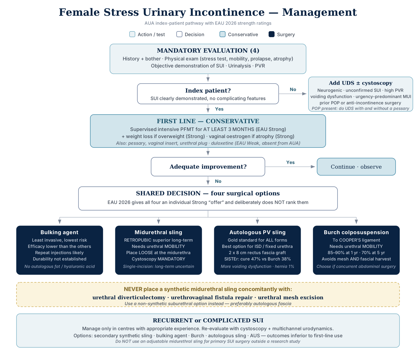
Click the algorithm to open it full size.
The 4 mandatory investigations
- History (including bother) and physical exam
- Bother is paramount to the decision to operate in the index patient
- Objective demonstration of SUI
- History alone does NOT diagnose SUI
- Inspection of pads soaked with urine is sufficient objective confirmation of leakage
- The demonstration does not have to happen in front of you
- Urinalysis
- PVR
Physical exam — 6 elements
- Stress test, supine and/or standing, comfortably full bladder
- Negative supine → repeat standing
- Symptomatic SUI but no leak on post-void exam → do not treat invasively until SUI is documented:
- Repeat a full-bladder stress test supine, then standing if necessary → still negative → offer UDS
- Symptomatic SUI but no leak on post-void exam → do not treat invasively until SUI is documented:
- Negative supine → repeat standing
- Focused abdominal exam
- Urethral mobility
- Q-tip test does not diagnose SUI
- SUI exists without hypermobility and vice versa; it only quantifies mobility.
- Hypermobility = >30° from horizontal
- Q-tip test does not diagnose SUI
- Pelvic prolapse
- Vaginal atrophy / estrogenization
- Focused neurologic exam
- Focused neuro-urological exam is routine in the initial evaluation of women with LUTS, including mixed incontinence
- Alongside a complete history, focused physical exam and cough stress test
- Focused neuro-urological exam is routine in the initial evaluation of women with LUTS, including mixed incontinence
- Pad test confirms incontinence but not the type
- Short test >1.4 g = incontinence
- 24-hour test >1.3 g = positive (some accept up to 8 g in 24 h as normal)
- 24-h severity 4.4–20 g mild · 21–74 g moderate · >75 g severe
- Assess frailty in patients ≥65 — distinct from comorbidity, predicts poor post-op outcomes
- Only 18–50% of women with pelvic floor disorders seek care — ask directly
- Voiding diary: 3 days is optimal, but a single day is acceptable
- Validated questionnaires: the ICIQ family — ICIQ-UI, ICIQ-SF, ICIQ-FLUTS, ICIQ-MLUTS
Cystoscopy — 3 indications
- Not in the index patient
- Suspected bladder pathology (e.g. microhematuria)
- Structural lower tract abnormality
- Undergoing MUS or PVS — confirm integrity, exclude foreign body
- Also: prior anti-incontinence or POP surgery if perforation is suspected
- Suspect it with new LUTS, hematuria or recurrent UTI
Urodynamics
- May be OMITTED in the index patient when SUI is clearly demonstrated
- VALUE (NEJM 2012): 630 women with uncomplicated SUI — office evaluation alone was NOT INFERIOR to evaluation plus UDS
- Uncomplicated SUI =
- Demonstrable SUI on exam
- No OAB symptoms
- Normal bladder emptying
- No prior SUI surgery
- No features suggesting a neurologic cause
- Basic office evaluation = pelvic exam with a full-bladder supine stress test · PVR · urinalysis → preoperative UDS did not improve surgical outcomes over it
- Uncomplicated SUI =
- 9 non-index triggers:
- Neurogenic LUTD
- Unconfirmed SUI
- Subjective/objective mismatch
- Elevated PVR
- Significant voiding dysfunction
- Significant urgency/UUI/OAB
- Abnormal urinalysis — unexplained hematuria or pyuria
- Prior POP surgery
- Prior anti-incontinence surgery
- Significant POP → UDS with and without a pessary
- Suspected SUI not seen on UDS, no prolapse → repeat stress testing with the urethral catheter removed
- Downsizing the catheter or removing all catheters is not recommended
- Prior pelvic radiotherapy = complicated SUI → filling cystometry before any sling
- Detects a small, poorly compliant, high-pressure bladder, where outlet surgery risks retention and upper tract deterioration
- Continuous leakage ("all the time"), not classically stress- or urge-related → a positive cough stress test is not enough to diagnose → UDS before any sling (no fistula risk factors)
- Bothersome MUI refractory to antimuscarinics → UDS before invasive therapy
- The findings, including pressure-flow emptying, decide between sling or bulking and sacral neuromodulation or onabotulinumtoxinA
- DO present → consider something other than a sling alone; small capacity and low compliance also worsen sling outcomes
- UDS answers two questions:
- (1) is this a storage problem only, or is there an emptying problem too?
- (2) is the incontinence a detrusor problem or an outlet problem?
- For higher-risk interventions — that is, surgery — multichannel UDS remains the most accurate test available, and is most useful after previous surgical intervention
Female SUI — Conservative & Medical
- Non-surgical options (4):
- Urethral plugs
- Vaginal inserts
- Continence pessary — chiefly for prolapse-related SUI
- PFMT ± biofeedback
- Observe if minimal bother, or not a candidate for other therapy
- No stem cell therapy outside trials
- Surgery only after conservative management has failed
Intravaginal continence devices — pessaries, tampons, contraceptive diaphragms; satisfaction >50%. But EAU does NOT recommend offering mechanical devices routinely (Strong) — the trade-offs below are why.
| Advantages | Disadvantages |
|---|---|
| Work across a broad spectrum of stress or mixed UI | Insertion and removal for cleaning can be difficult |
| Fitted in clinic without invasive testing | Vaginal discharge, odor, irritation |
| Worn only for predictable activities that provoke leakage | Dislodgement |
| Mild side-effect profile | Cannot address ISD, and cannot transmit pressure properly to a fibrotic urethra |
| Also manage bothersome prolapse symptoms |
- Situational SUI in a young woman (leaks only during sports) despite regimented PFMT → incontinence pessary or device
- Used only for short periods
- Defer slings until after childbearing if possible — likely excessive here.
- Radiofrequency ablation is invasive and not well tested in young women
- Urinary diversion is a last resort — refractory cases with severe sphincter damage only
- Bladder neck closure with simultaneous lower-tract reconstruction can succeed when the outlet is destroyed
- A functional urethral closure with a pubovaginal sling leaves a pop-off valve at high pressures and preserves transurethral access
| Intervention | Position |
|---|---|
| Supervised intensive PFMT ≥3 months | First line for all SUI and MUI (Strong) — including elderly and pre/postnatal |
| Weight loss | Encourage in all overweight/obese (Strong) |
| Vaginal estrogen | Offer in postmenopausal women with vulvovaginal atrophy (Strong) |
| Electrical stimulation | Consider, or as an adjunct for teaching a contraction (Weak) |
| Electroacupuncture | Offer (Strong) |
| Electromagnetic stimulation | Do NOT offer outside research (Strong) |
| Vaginal laser | Do NOT offer outside research (Strong) |
| Duloxetine | Offer to selected patients unresponsive to conservative therapy who want to avoid surgery (Weak); titrate on and off (Strong) |
PFMT
- PFMT evidence: 8× more likely to report cure; as effective as surgery for about half of women
- Biofeedback adds nothing when dose and attention are matched; group training is non-inferior and cost-effective
- Less effective when MUI/UUI patients are included; more effective if intensive and supervised
- Head-to-head vs MUS at 12 months: improvement 90.8% surgery vs 64.4% physiotherapy
- But 49% of the physiotherapy arm crossed over to surgery
- Uncomplicated SUI
- Leaks on cough, normal POP-Q (all points negative, short of the hymen), can contract her pelvic floor → start PFMT
- UDS unnecessary before non-invasive therapy; biofeedback may not be required when she can already contract
- Postpartum SUI persisting >3 months → pelvic floor muscle therapy first; UDS not needed when the leak is demonstrable
- Bulking agents and slings (MUS or autologous) only after conservative failure — ideally after childbearing
Drugs
- Duloxetine mechanism: blocks presynaptic 5-HT and norepinephrine reuptake → enhanced pudendal motor neuron drive → increased sphincter resting tone
- Cure ≈10%
- Do NOT offer antimuscarinics when the problem is primarily SUI — they treat the wrong mechanism
- α-agonists raise urethral muscle tone, but the evidence that they beat placebo is weak
- TCAs (imipramine) are the other oral option alongside the SNRI duloxetine
- But imipramine is off-label for SUI, has potentially harmful side effects, and is not included in the AUA/SUFU guideline
Estrogen
- Oral estrogen ± progestogen makes incontinence WORSE
- It increases the incidence of SUI and UUI in women continent at baseline and worsens UI in those already incontinent.
- This is the opposite of vaginal estrogen, which treats vulvovaginal atrophy and is offered (Strong)
- The risk applies to current OR past systemic hormones — oral estrogen or birth control pills — in middle-aged and older women
- Topical estrogen does not raise it
- Low endogenous estrogen levels do NOT increase SUI risk
- Vaginal estrogen is preferred to systemic therapy for LUTS
- Helps the genitourinary syndrome of menopause in atrophic postmenopausal women (dryness, itching, urgency and frequency — OAB symptoms), urgency incontinence and recurrent UTIs
- Not an effective treatment for SUI, so not routine SUI therapy
- Apart from urgency incontinence, it has not been shown to treat urinary incontinence
- Mechanism (vaginal or oral estrogen):
- More urethral α-adrenergic receptors
- Thicker urothelial mucosa → better urethral seal
- Increased blood flow.
- Vaginal acidification helps UTIs, not continence
Female SUI — Surgery
The 4 options
- Periurethral bulking agent
- Midurethral sling (MUS)
- Autologous pubovaginal sling (PVS)
- Burch colposuspension
All four carry an individual Strong "offer" — they are deliberately not ranked. Choose by shared decision-making.
- Anterior colporrhaphy alone must NOT be used to treat SUI (Grade A)
- Combine it with a sling.
- Needle suspensions are abandoned
Bulking agents
- Bulkamid (polyacrylamide hydrogel), FDA approved 2020; little long-term data
- Indications (2):
- Wants to avoid more invasive surgery / longer recovery
- Insufficient improvement after a previous anti-incontinence procedure
- Counsel on the expected need for repeat injections
- Ideal candidate:
- Isolated ISD
- ALPP <100 cm H₂O
- Minimal hypermobility (≤15° descensus)
- Inject at the PROXIMAL urethra
- Too near the bladder neck → extravasation and poor coaptation
- Too distal into the sphincter → failure, pain, irritative symptoms
- Transurethral: 3 and 9 o'clock · periurethral: 4 and 8 o'clock
- Efficacy up to 75% improvement short-term, substantially less at 1 year — improvement ≠ dryness
- AEs:
- Pain at implantation 13%
- Retention 6%
- UTI 4%
- Hematuria 2%
- Do NOT use autologous fat or hyaluronic acid (Strong)
- Higher AE risk.
- Collagen is no longer available
- Agents used historically:
- Collagen
- PTFE
- Dextranomer/hyaluronic acid
- Durasphere (graphite-coated beads)
- Coaptite (calcium hydroxyapatite)
- Polydimethylsiloxane elastomer (Macroplastique)
- All share a short duration of effect, which is why repeat injection is the expectation
Midurethral sling
- Three types:
- Retropubic (TVT-R)
- Transobturator (TVT-O)
- Single-incision
- Mechanism (2):
- Restricts posterior urethral wall movement
- Narrows the lumen as the posterior wall moves inward
- Material: macroporous monofilament polypropylene, pore size >75 μm
- Allows ingrowth, decreases encapsulation and infection
- Patients without urethral hypermobility respond poorly — a fixed urethra means ISD
- Exclude POP and a urethral diverticulum before offering an MUS — both change the operation
NEVER place a synthetic MUS concomitantly with: urethral diverticulectomy · urethrovaginal fistula repair · urethral mesh excision. Use autologous fascia.
- Also avoid mesh where healing is poor:
- Prior radiation
- Significant scarring
- Poor tissue quality
- Long-term steroids
- Impaired collagen from systemic autoimmune disease (Sjögren's, SLE)
- Immunosuppression
- Tension: place LOOSE at the midurethra
- A loose sling with a mobile urethra compresses on Valsalva but is non-obstructive at rest
- Single-incision slings are the exception — tension them TIGHTER, and restriction of mobility afterwards predicts a better result
- Cystoscopy is mandatory (Strong)
- Perforation → withdraw and repass more laterally, catheter 5–7 days
- Exam answer: TVT bladder perforation recognized intraoperatively → withdraw, re-pass more laterally, complete the sling, then Foley 3–7 days
- Trocar in the URETHRA is different → repair the urethrotomy, ABORT the procedure, indwelling catheter
- Never place a mesh sling after an inadvertent urethral injury (AUA/SUFU)
- Retropubic anatomy:
- Obturator vessels are the closest major vessels
- Dorsal nerves of the clitoris run on the inferior surface of the ischiopubic rami, ~1.4 cm from the midline
- Keep the needle on the bone
- Transobturator anatomy: avoids the true pelvis.
- Traverses: obturator internus → obturator membrane → obturator externus → adductor magnus, adductor brevis, gracilis.
- Must NOT traverse: adductor longus or levator ani
- Does NOT pass through the rectus fascia (the retropubic sling does)
- Medially the trocar enters the anterior compartment of the ischiorectal fascia
- Nerves running close to the pass: obturator nerve, dorsal nerve of the clitoris, pudendal nerve
- Avoid the adductor longus tendon — contact causes substantial pain
- Stay medial and close to bone — obturator nerve and vessels lie cephalolateral in the canal
- Skin entry at the notch where the adductor longus tendon meets the inferior pubic ramus, at the level of the clitoris
- Urethral injury on dissection → repair 4-0 over a 16F catheter and POSTPONE the sling 6–12 weeks
Outcomes
- Retropubic and transobturator look similar short-term, but retropubic is SUPERIOR long-term (Strong)
- ESTER OR 0.74, SUCRA 89.1% vs 64.1%
- TOMUS long-term:
- Transobturator less effective for continence
- Retropubic → higher PVRs, more recurrent UTIs, more delayed sling erosion into the bladder
- Single-incision slings lose efficacy with longer follow-up — short-term equivalent, long-term uncertain
- ISD: success is lower with a fixed urethra and low LPP
- Retropubic > transobturator in ISD.
- Low LPP alone is not a contraindication
- Pre-operative urethral mobility predicts success
- Not compromised by concomitant vaginal surgery, age, or obesity
- Elderly: cure comparable, but more persistent SUI and more de novo urgency; do not exclude on age alone
- Elderly detail:
- The de novo rise covers urgency AND urgency incontinence after any sling material, MUS included
- The comparable cure holds with urethral hypermobility, and the extra persistent SUI is possibly from coexisting mixed incontinence or less urethral hypermobility
- Obese: higher bladder trocar injury in NON-obese patients
- Recurrent SUI:
- Salvage efficacy ≈ primary
- Higher bladder perforation risk after prior retropubic suspension
Autologous pubovaginal sling
- Gold standard for ALL forms of SUI
- Indications:
- ISD (retropubic suspension is contraindicated in ISD)
- Urethral hypermobility
- Deficiency of the midurethral complex
- MUI
- Concomitant cystocele
- Urethral diverticulum
- Neuropathic bladder — e.g. myelodysplasia, to treat the SUI that emerges once CIC begins
- After trauma
- After failed retropubic suspension or failed MUS
- After mesh erosion
- Urethral reconstruction
- Also:
- Proximal urethral loss from a long-standing indwelling catheter
- Refractory SUI after failed MUS and bulking agents
- Prior anti-incontinence surgery + fixed "pipe-stem" urethra + low Valsalva LPP = ISD → autologous PVS rather than another MUS
- NOT indicated: low urethral resistance with poor bladder compliance
- The incompetent outlet is acting as a pop-off, so closing it raises storage pressure → correct compliance first (antimuscarinics · onabotulinumtoxinA · augmentation), then treat the outlet
- Graft: 2 cm × 8 cm rectus fascia (most common) or fascia lata (classically 20 × 2 cm, now often 8 cm)
- Keep the transverse fascial incision ≥2 cm from the symphysis for tension-free closure
- Positioned at the bladder neck / proximal urethra
- Via an inverted-U vaginal incision
- Through a combined abdominal and transvaginal approach
- Both ends end up in the retropubic space
- Key bony landmark: the ischiopubic (pubic) rami
- Perforate the endopelvic fascia just under the palpable ramus, scissors directed superolaterally toward the ipsilateral shoulder
- Drain the bladder before fascial perforation and needle passage → cystoscopy with the needles in place
- Bladder perforation → withdraw and repass more laterally
- Tension: tie above the rectus fascia leaving a TWO-FINGERBREADTH gap.
- Close the vagina, remove the speculum, and finish any POP repair before final tensioning
- Advantages:
- Minimal inflammation
- Negligible urethral erosion risk
- Disadvantages:
- Longer operation and stay
- Post-op pain
- Suprapubic seroma
- Incisional hernia (~1%)
- Synthetic PVS is no longer used — inflammation, infection, perforation, exposure
| Graft | Source | Note |
|---|---|---|
| Autologous | The patient's own tissue | Best for PVS — negligible risk of urethral erosion |
| Allograft | Another human | |
| Xenograft | Animal | |
| Synthetic | Manufactured | No longer used in PVS |
- Autologous or allograft sling perforating the urethra:
- Incise or excise the intraluminal portion only and close the urethra simply.
- Additional coverage such as a Martius flap is rarely needed — unlike a synthetic sling, which almost always needs it
| PVS | MUS | |
|---|---|---|
| Position | Bladder neck | Midurethra |
| Material | Autologous | Synthetic |
| Donor morbidity | Yes | None |
| Corrects | ISD and hypermobility | Hypermobility only |
| With diverticular repair | Can be used | Cannot be used |
| Voiding dysfunction | Higher | Lower |
| Retention | Higher risk — tensioned at the bladder neck | Least characteristic complication — placed loosely at the midurethra |
- SISTEr (NEJM 2007): PVS vs Burch
- Cure 47% vs 38%, but significantly more voiding dysfunction and cystitis
- All 20 BOO procedures were in the PVS group
- 5 years: continence 31% vs 24%, satisfaction 83% vs 73%; serious AEs similar (9–13% vs 10%)
- 33% have persistent urge incontinence and up to 9% develop de novo UUI
Burch colposuspension
- Indications (2): wants to avoid mesh AND avoid fascial harvest · undergoing a simultaneous abdominal procedure
- Also when vaginal access is limited (contractures, stenosis)
- Concomitant abdominal surgery that cannot be done vaginally
- E.g. SUI + planned open ureteric reimplantation → Burch: retropubic space already open, no implant added.
- Several RCTs: outcomes essentially equivalent to TVT
- Frequently high intra-abdominal pressure (chronic cough, strenuous work)
- Retropubic repair is considered, though a PVS may serve these patients better
- Requires urethral mobility
- And adequate vaginal mobility and capacity
- The lateral vaginal fornices must reach Cooper's ligament on each side → hence contraindication 4
- And adequate vaginal mobility and capacity
Contraindications (4)
- Prior failed incontinence surgery with a high likelihood of ISD
- SUI due solely to ISD — a fixed, non-functional proximal urethra
- Pan-pelvic floor weakness — needs a combined pelvic floor repair instead, especially with a central cystocele, rectocele or introital deficiency
- Inadequate vaginal length or mobility — after previous vaginal surgery or radiotherapy
- Elevates the anterior vaginal wall and paravesical tissue toward Cooper's (iliopectineal) ligament with 2–4 sutures of the pelvic sidewall
- The urethral wall is deliberately NOT included.
- Sutures pass medial-to-lateral
- Tie distal first with knots anterior to the ligament, aiming for two fingerbreadths between bladder neck and pubic bone
- A V-shaped suspension increases obstructive problems — and the repair remains suture-dependent long term
- Be ready for sudden copious bleeding at Cooper's ligament
- Success 85–90% at 1 year, 70% at 5 years; laparoscopic = open
- Failure predictors:
- BMI >25
- Anticholinergic use
- Age >60
- Prior SUI procedure
- Previous surgery is the most important determinant of retropubic outcome
- Burch success 81% after one failed repair → 25% after two → 0% after three
Two complications specific to colposuspension
- Enterocele — 5%.
- Commoner after Burch because it aggravates posterior wall weakness.
- Prophylactic obliteration of the cul-de-sac reduces the risk
- De novo posterior or apical prolapse overall: 2.5–27%.
- Mechanism = altered vaginal axis
- Lifting the anterolateral vagina to Cooper's ligament pulls the proximal vagina forward and up, so the posterior cul-de-sac is exposed to intra-abdominal pressure
- Mechanism = altered vaginal axis
- Formally assess the posterior and apical compartments (POP-Q or Baden-Walker) before and after any retropubic suspension
- Fix coexisting or occult defects at the same sitting — e.g. culdoplasty or apical suspension for an already deep cul-de-sac
- Post-colposuspension syndrome — pain in one or both groins at the suspension site
The other retropubic operations
| Operation | Fixed to | Technique | Position |
|---|---|---|---|
| MMK | Periosteum of the symphysis pubis | 3 sutures at the bladder neck — involves the urethral and vaginal wall, unlike Burch | No longer recommended. Osteitis pubis 1–3%; suture-dependent; less durable than Burch |
| Paravaginal repair | ATFP | 3–8 sutures through the lateral vaginal sulcus and overlying fascia. Bladder and urethra are not mobilized off their vaginal attachments | Not recommended for SUI alone (Grade A); less effective than other colposuspensions. Horizontal suspension → obstruction is less common |
| VOS (vagino-obturator shelf) | Internal obturator fascia ± the iliopectineal line as a modification | Anchors the vagina laterally | Horizontal suspension → obstruction is less common |
| Needle suspensions | — | — | Abandoned — suture pull-through |
- There is a laparoscopic version of all of them
- Retropubic beats the vaginal route (anterior colporrhaphy) for long-term results, and is the better choice with a concomitant cystocele — where it should be combined with a sling
AUS in women
- Second-line.
- Continence 79.6%, but explantation 13% · revision 15% · vaginal erosion 9% · mechanical failure 13%
- Transvaginal implantation is not favored (infection, cuff erosion)
- Counsel: high risk of complications, mechanical failure or explantation even in expert centers (Strong)
SUI Surgery — Complications
MUS — the 10 to quote
| Complication | Rate |
|---|---|
| Bladder trocar injury | 2.7–3.8% overall — retropubic 2.7–23.8% vs transobturator 0–1.3% |
| Voiding dysfunction | 7.6% |
| Vaginal mesh exposure | 0.5–8.1% (up to 9%) |
| Mesh perforation of bladder | 0.5–0.6% |
| Wound healing problems | 1% |
| Bleeding / vascular injury | Higher with retropubic; severe bleeding 2–3% |
| Groin/thigh pain and numbness | 10–15% with transobturator |
| Sexual dysfunction · infection · bowel perforation · death | Rare |
- Trocar injury is generally a benign condition
- Bladder injury is the least likely TOT complication
- The trocar crosses obturator internus, obturator membrane and obturator externus and never enters the retropubic space
- Bladder injury is the least likely TOT complication
- Vaginal exposure:
- Manifests weeks to months post-op.
- Risk factors
- Diabetes
- Smoking
- Older age
- Previous vaginal surgery.
- Two more risk factors: vaginal incision >2 cm · poor vaginal estrogenization — so topical estrogen treats a cause, not just the defect
- Observation is only for the asymptomatic woman with an exposure <1 cm, with estrogen ± antibiotic cream.
- Otherwise: vaginal advancement flap or excision via a transvaginal approach
- <1 cm exposure of a large-pore monofilament sling → topical estrogen and re-evaluate in several weeks — not explantation.
- Excise the exposed segment for failed conservative therapy, a larger defect or bothersome dyspareunia
- Why small exposures heal: large-pore monofilament mesh allows fibroblast in-growth and macrophage intercalation.
- Timing of primary wound closure is controversial; drains are not recommended
- Other conservative routes: topical estrogen with delayed primary closure, or healing by secondary intention
- Exam answer: an asymptomatic extrusion that bothers neither her nor her partner (e.g. 1 cm, found incidentally years later) → can be observed, continuing estrogen if she has atrophy.
- Excise only for bother — unnecessary excision risks recurrent SUI
- Urethral perforation → observation is NEVER acceptable.
- Transurethral excision of the tape with closure of the urethrotomy ± Martius flap
- An autologous PVS may be placed at the same sitting.
- Endoscopic tape transection has a role for a small intraluminal segment
- 42% develop SUI after intraurethral mesh excision — a same-sitting autologous fascial PVS significantly lowers that risk
- Mesh perforating the BLADDER → never observe this either.
- Fill the bladder completely before you look
- An underfilled bladder hides the mesh in a fold, and that is the commonest reason it is missed.
- Send a urine culture and consider upper tract imaging to assess the ureters.
- First: endoscopic excision.
- If that fails: a combined transvaginal and retropubic approach
- Suspect urethral or bladder perforation when new storage symptoms, recurrent UTIs and microscopic hematuria appear months after a MUS → cystoscopy
- Mesh found in the lumen → excise it
- Endoscopic bladder management works only with absolute excision of ALL exposed mesh
- Fill the bladder completely before you look
- Exposures and perforations are clearly linked to technique and errant sling placement.
- Management is individualized, but all exposed sling material — vaginal or within the urinary tract — must end up completely excised or covered (conservative healing counts as cover)
- Pain:
- Most groin pain settles by day 2; discomfort up to 2 weeks.
- Refractory pain → exclude extrusion, organ injury, adductor longus perforation.
- Localized pain → limited urethrolysis
- Widespread pain or infection → complete excision.
- Removal does not guarantee pain relief
- Transobturator leg pain is commoner with the inside-out pass and in athletic, muscular builds.
- Escalation if it does not settle: pain service → sling removal
- TOMUS: transobturator slings caused more neurologic complications than retropubic
- Injury to or compression of small pudendal or obturator nerve branches
- Voiding dysfunction after MUS:
- Usually obstruction — sling too tight, too proximal, or associated POP.
- Rule out POP and do cystoscopy.
- Approach her as a new patient and manage conservatively first
- No preoperative factor predicts it
- Not every case is obstruction:
- New frequency and urgency after TVT with a normal flow (e.g. Qmax 18 mL/s) = transient de novo urgency (up to 12%) → treat the storage symptoms
- Early retention is usually temporary → start CIC
- Hematuria or pyuria → CIC + cystoscopy before any incision, to rule out urethral or bladder mesh perforation.
- If symptoms began with the sling, incision is likely needed whatever the UDS shows
- Hesitancy, UUI, staccato flow and a raised PVR (e.g. 150 mL) → UDS first to separate obstruction from detrusor underactivity:
- Confirmed obstruction → incise
- Confirmed DU → CIC.
- Avoid antimuscarinics with incomplete emptying
- Wait 2–4 weeks before considering sling release
- Exam answer: conservative management for a few days to 1 month, then contemplate release when voiding trials persistently fail — immediate release is not recommended (a short wait usually resolves it)
- In frank retention, intervene sooner — within 4 weeks — because it is unlikely to resolve
- Within 3 months → the sling can be LOOSENED:
- Reopen the vaginal incision, pass an instrument around the mesh and tug.
- If it is embedded and will not loosen, simply DIVIDE it — the entire sling does not need to be excised
- At this stage, save excision for urethral erosion — as a release it may raise the recurrent SUI risk over a midline incision alone
- After 3 months → the sling is fixed along its whole course.
- A midline incision may not release enough — go to formal sling excision and urethrolysis
- Urethral dilatation is ineffective in obstruction and risks urethral extrusion of the mesh
- Dilation is contraindicated after a mesh sling
- It traumatizes the tissue over the inflexible mesh.
- Tamsulosin will not relieve an anatomic obstruction either
- Retropubic worse than transobturator: surgery-requiring voiding dysfunction 2.7% vs 0%, retention >6 weeks 3.7% vs 0.7%
- MUS infection → treat conservatively first; remove the sling if that fails
PVS obstruction and urethrolysis
- Transient retention is common — most void spontaneously within 10 days
- Voiding dysfunction after PVS 2.5–35% · retention beyond 4 weeks ~8% · permanent retention generally ≤5% — far more than after a loosely placed MUS
- If she develops UUI or obstruction after a PVS, work her up as a new patient
- History, examination, urinalysis, PVR, videourodynamics and cystoscopy
- Conservative package:
- Indwelling catheter or CIC
- Obstruction after a fascial sling → CIC first — fascial slings loosen over time, and serial PVRs track the improvement
- Timed voiding
- Double voiding
- Biofeedback
- PFMT
- α-blockers ± anticholinergics
- Indwelling catheter or CIC
- Within 6 weeks: loosen the sling in theatre
- Cystoscope in, gentle caudal traction on the urethra.
- Not advised with synthetic slings
Complete obstruction — revision may be brought forward
- Loosen the sling under cystoscopic vision with gentle downward pressure on the urethra — usually ineffective, and contraindicated with a synthetic sling
- Urethral dilation with downward traction
- Transurethral resection or incision of the bladder neck — not recommended: it risks the sphincter and the bladder neck, and periurethral fibrosis can leave worse incontinence or a bladder neck contracture
- After 6 weeks, or when conservative measures fail → sling incision, then formal urethrolysis if that fails.
- Better to wait until 3 months post-operatively before a formal urethrolysis
- Exam answer: after an autologous fascial sling, sling incision waits 3–4 months — versus within the first month after a MUS
- Sling incision: comparable success, shorter, less morbid — but recurrent SUI 34% vs 0–19% after formal urethrolysis
- Approaches: retropubic · transvaginal · suprameatal; success 65–93%; no parameter predicts success
- The temporal relationship between symptoms and surgery is the key criterion — UDS is essential
- Failure of urethrolysis — 4 causes: persistent/recurrent obstruction · DO · impaired contractility · learned voiding dysfunction
- Most common reason is insufficient dissection → repeat urethrolysis is justified
- OAB is refractory in 50% after urethrolysis → consider SNM
- Always rule out apical, anterior and posterior prolapse as a cause of obstruction
Recurrent / complicated SUI
- Experienced centers only (Strong)
- Re-evaluate with cystoscopy and multichannel UDS
- Second-line surgery is inferior to first-line use — less effective, more complications (Weak)
- Options: secondary synthetic sling · bulking · Burch · autologous sling · AUS (Weak)
- Recurrent SUI after a MUS was removed, or excised for urethral erosion → autologous fascial PVS — avoids putting synthetic material back into a scarred bed
- Do NOT use an adjustable MUS for primary SUI outside research (Strong)
SUI Surgery at the Time of POP Surgery
The single biggest AUA/EAU divergence in this topic.
AUA
- Adding a retropubic MUS to fascial plication, Burch, or abdominal sacrocolpopexy may reduce post-operative SUI (Cochrane, 22 RCTs)
- CARE trial: prolapse without pre-op SUI, sacrocolpopexy ± Burch → significantly higher incontinence at every follow-up point without the Burch
- Vault prolapse without SUI symptoms → sacrocolpopexy alone ONLY if neither examination nor UDS shows SUI.
- Objective SUI on either → counsel for concomitant SUI surgery (CARE and OPUS trials)
- A prior anti-incontinence procedure (e.g. a bulking agent) does not rule out a concomitant sling
- Objective SUI → still counsel for SUI surgery at the prolapse repair
- Vault prolapse without SUI symptoms → sacrocolpopexy alone ONLY if neither examination nor UDS shows SUI.
- Trade-off: prolapse repair at the time of sling → more outlet obstruction (9.4% vs 5.5%) but fewer repeat SUI or POP procedures at 1 year
- MUS placed for urodynamic or occult SUI at POP repair:
- Risk of intervention for obstruction = risk of intervention for SUI without it (8.5% vs 8.3%)
EAU 2026
- Symptomatic or occult SUI:
- Offer simultaneous surgery ONLY after full discussion of risks vs POP surgery alone (Strong)
- Inform of increased AEs with combined surgery (Strong)
- No symptomatic or occult SUI:
- Inform of de novo SUI risk (Strong)
- DO NOT offer concomitant anti-incontinence surgery at ABDOMINAL prolapse surgery (Strong)
Both agree (Strong) that occult SUI must be tested for and discussed. The disagreement is only whether to act on it prophylactically at an abdominal repair.
Matching the concomitant repair to the POP-Q
- Post-hysterectomy anterior and apical prolapse (Ba +4), Bp −3, no SUI with the prolapse reduced → vault suspension + anterior repair
- No SUI on prolapse reduction → no absolute indication for an anti-incontinence procedure
- Bp −3 = intact posterior compartment → no posterior repair.
- Anterior repair alone → unacceptably high failure
- SUI + Ba +1, C 0, D −5, Bp −2 → sling + vaginal hysterectomy + cystocele repair
- C 0 = uterovaginal prolapse
- C 0 vs D −5 = elongated cervix → hysteropexy or sacrouteropexy would leave it unaddressed
- Ba +1 = anterior wall laxity → cystocele repair
- Bp −2 = no posterior laxity → no rectocele repair
- SUI + Aa/Ba −2, Ap/Bp +2, C 0 → MUS + posterior and apical repair (e.g. posterior colporrhaphy + sacrospinous fixation)
- Ap/Bp +2 = posterior wall beyond the hymen
- C 0 = apex at the hymen.
- Aa/Ba −2 = anterior wall supported → no anterior repair
- A posterior repair alone misses the apex
Urogenital Fistula — Principles
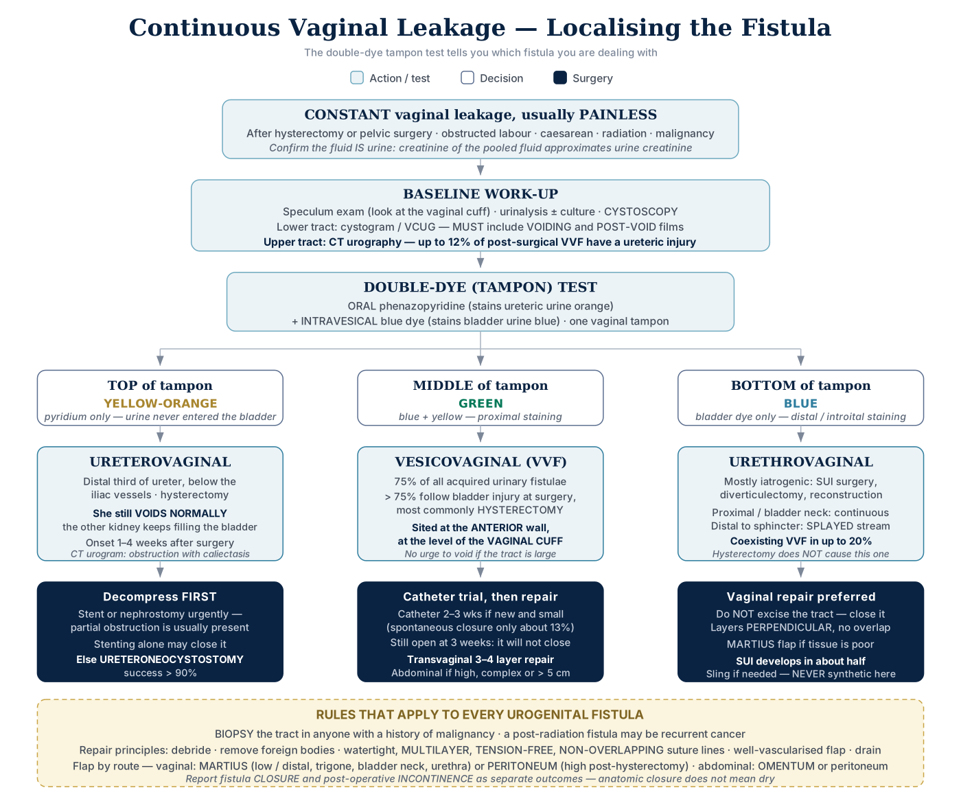
Click the algorithm to open it full size.
- Fistula = extra-anatomic communication between two epithelial- or mesothelial-lined cavities or the skin
"Fistula TO MIIND" — 8 factors
| Factor | |
|---|---|
| F | Foreign body |
| T | Technical surgical problems |
| O | Obstruction — remove or bypass distal obstruction |
| M | Malignancy — biopsy the tract in anyone with prior malignancy |
| I | Ischemia |
| I | Infection |
| N | Nutrition |
| D | Drainage — unobstructed drainage ± stenting |
- Leak persisting after a repaired injury in a cancer patient → biopsy before any repair
- A bladder leak still present 9 weeks after repairing a bladder injury at colon cancer surgery → cystoscopy + biopsy to exclude recurrent or persistent malignancy before definitive repair
- Further catheter drainage and a repeat cystogram add little by then — think foreign body, inadequate drainage or malignancy
11 principles of repair
- Adequate exposure
- Debride devitalized and ischemic tissue
- Remove foreign bodies / synthetic material
- Separate the involved cavities
- Watertight closure
- Well-vascularized healthy tissue flaps, atraumatic handling
- Multiple layers
- Tension-free, NON-OVERLAPPING suture lines
- Adequate drainage ± stenting
- Treat and prevent infection
- Hemostasis
- Excising the epithelialized fistula tract is NOT a required principle
- It may help, but is not needed in every repair
- Tension-free watertight closure, vascularized flaps and adequate drainage are the core
5 factors that prevent VVF at gynecological surgery
- Immediate detection of bladder injury, using dyes if necessary
- Watertight closure of the bladder
- Satisfactory extravesical drain placement
- Avoid a vaginal incision if possible once a bladder injury has been recognized
- Prolonged, uninterrupted post-operative bladder drainage
Vesicovaginal Fistula
- 75% of acquired urinary tract fistulae
Causes — "Radical Obstetrical Colleagues' Trauma Causes Incontinence Fistula"
| Cause | |
|---|---|
| R | Radiation — may appear decades later; any post-radiation fistula may be recurrent malignancy |
| O | Obstetric |
| C | Cancer |
| T | Trauma |
| C | Congenital |
| I | Infectious / inflammatory |
| F | Foreign body (vaginal prolapse mesh) |
| + | Post-surgical — dominant in the industrialized world |
- Radiation fistula — timing tells the mechanism:
- During or shortly after radiotherapy → tumor necrosis in the bladder or vaginal wall
- ≥ 1 year later → radiation endarteritis obliterans → necrosis of the bladder and vaginal wall
- Developing world: prolonged obstructed labor → pressure necrosis.
- Young primigravida with a narrow pelvis
- Part of the obstructed labor injury complex
- Obstetric fistulae are LARGER, more DISTAL, and involve the BLADDER NECK and PROXIMAL URETHRA → difficult to repair
- Industrialized world: >75% follow surgical bladder injury — most commonly HYSTERECTOMY
- Bladder injury at abdominal hysterectomy 0.5–1.0%; fistula after hysterectomy 0.1–0.2%
- Of the surgical cases:
- Hysterectomy for benign disease 60–75%
- Hysterectomy for malignancy ~30%
- Cesarean section ~6%
- Obstetric injury ~1%
- (approximate shares — they do not sum cleanly)
- Three mechanisms:
- (1) unrecognized cystotomy near the vaginal cuff → urinoma → drains through the cuff → epithelialized tract
- (2) tissue necrosis from cautery
- (3) a suture placed through both bladder and vaginal wall at cuff closure, or a hemostatic ligature taken blindly for pelvic bleeding
- Urogenital fistula is ≈9× more likely after radical hysterectomy for malignancy than simple hysterectomy
- Clear vaginal discharge after hysterectomy is not always urine
- Also seroma, lymphatic fistula, peritoneal fluid, peritoneovaginal fistula, normal vaginal secretions, tubal fluid, vaginitis
Post-operative risk factors
| General | Specific |
|---|---|
| Diabetes | Malignancy |
| Ischemia | Neurogenic bladder |
| Arteriosclerosis | Cesarean section / prior uterine surgery |
| PID or infection | Pessary use |
| Prior radiotherapy | Endometriosis |
| Steroids | Endocervical conization (scarring) |
Classification
What matters clinically — simple vs complicated
| Simple | Complicated |
|---|---|
| ≤ 3 cm | > 3 cm in diameter |
| First repair | Recurrent, or a failed previous repair |
| No radiotherapy | Prior radiotherapy |
| Benign cause | Associated with malignancy |
| Away from the trigone and urethra | At the trigone, bladder neck or urethra |
| Healthy tissue | Compromised tissue that will heal badly |
- Formal classification systems (brief): WHO — simple vs complex (4 cm cut-off; sphincter, ureteric or circumferential involvement makes it complex) · Goh — by distance of the distal edge from the meatus (type 1 > 3.5 cm → type 4 < 1.5 cm; the better predictor) · Waaldijk — by involvement of the closing mechanism (I / IIA / IIB / III). In each, a higher type means worse closure and more incontinence
Diagnosis
- Most common complaint: CONSTANT vaginal drainage.
- Pain is uncommon
- Pain, when present → most likely a wound infection
- Also recurrent cystitis and perineal skin irritation
- Timing: either early — when the catheter comes out after the index operation — or at 1–3 weeks.
Pelvic exam
- Always do a speculum exam — post-hysterectomy VVF sits on the anterior vaginal wall at the vaginal cuff
- Assess surrounding inflammation — it determines the timing of repair
- Record vaginal depth, coexisting prolapse, atrophy and introital size — each of them changes the surgical approach
- Palpate for pelvic masses that may need dealing with at the same operation, and look for postmenopausal atrophy — she may need pre-operative estrogen
- Look for available flaps — ask about previous perineal, lower abdominal or thigh surgery, which may have used the tissue you were counting on
Fluid creatinine
- Confirm the fluid is urine: creatinine of the pooled fluid ≈ urine creatinine
- Fluid creatinine ≈ serum creatinine → peritoneovaginal fistula (peritoneal fluid, not urine)
Cystoscopy and cystogram
- Cystoscopy with retrograde bladder filling using a colored fluid confirms the diagnosis (Weak)
- And a VVF close to a ureteric orifice may force an abdominal approach with reimplantation, which is a decision to make at cystoscopy, not in theatre
- Cystoscopy is MANDATORY before repair
- Sensitivity 92–93%
- Defines fistula number, size and location relative to the ureteric orifices and bladder neck
- Assesses bladder capacity, and allows biopsy of any suspicious area
- A cystogram without VOIDING or POST-VOID images is NON-DIAGNOSTIC
- And take it in the LATERAL position, so that bladder and vagina do not overlap
Upper tract
- Image the upper tract
- Up to 12% of post-surgical VVFs have a ureteric injury or ureterovaginal fistula
- Mandatory, and done together with cystoscopy (≈ 10–12% coexist)
- Cystoscopy under anesthesia allows retrograde pyelography and ureteric stenting at the same sitting
Biopsy
- Biopsy the defect in anyone with malignancy or radiation history
- Includes suspected malignancy
- Even a post-radiotherapy cervical cancer fistula with a normal CT
- Because tumor in the tract precludes local repair (→ staging and cancer therapy, often urinary diversion)
- Includes suspected malignancy
The dye tests
Single dye (intravesical blue), vaginal packing
| Staining | Diagnosis |
|---|---|
| Introital / distal | Incontinence or urethrovaginal fistula |
| Proximal | VVF |
| Dye-free → repeat with IV indigo carmine → proximal staining | Ureterovaginal fistula |
- Bladder filled with up to 300 mL methylene blue; mid-tampon blue also → VVF; the incontinence behind distal staining is SUI
Double-dye (tampon): oral phenazopyridine + intravesical blue
| Tampon segment | Color | Diagnosis |
|---|---|---|
| Top | Yellow-orange | Ureterovaginal |
| Middle | Green | VVF |
| Bottom | Blue | Urethrovaginal |
| No stain anywhere | Clear | Peritoneovaginal fistula |
- Best test to localize a post-hysterectomy fistula
- Oral phenazopyridine (100–200 mg) + intravesical methylene blue: orange → ureterovaginal, blue → VVF
- Why orange = ureter:
- Phenazopyridine is renally excreted, so it reaches the vagina only from above the bladder
- Orange on the proximal swab only → ureterovaginal fistula
- Why the middle turns green: bladder blue mixes with yellow-orange phenazopyridine in that segment
- Orange top · dry middle · faint blue bottom → ureterovaginal fistula + urethral leakage
- Blue that skips the middle and shows only distally has left through the urethra, not a bladder–vagina tract
- Oral phenazopyridine (Pyridium) alone → an orange pad confirms the leak is urine, but not where the fistula is
- Pad stays clear after Pyridium following hysterectomy → the watery leak is not urine → peritoneovaginal fistula
- Methylene blue + indigo carmine given together cannot localize
- Both dyes are blue, so a bladder fistula cannot be told from a ureteric one
- A negative dye test may just be a dry moment — have her walk for a while and repeat it
Management — 4 options
1. Indwelling catheter
- Trial 2–3 weeks of catheter drainage with an antibiotic and an anticholinergic in newly diagnosed VVF
- Spontaneous closure ≈13% — essentially only small fistulae under 5 mm
- Start drainage immediately to prevent epithelialization
- Most favorable: <2–3 mm, simple injuries without devascularization or thermal spread
- Still open at 3 weeks → it will not close
- Classic candidate: small, newly diagnosed, post-hysterectomy VVF in non-irradiated tissue → urethral catheter trial
- Not healed → early vaginal repair
- Dry with the catheter in → keep draining; still leaking despite the catheter → spontaneous healing unlikely
- Nephrostomies or a suprapubic cystostomy add nothing over urethral catheter drainage
2. Fulguration — for small epithelialized tracts <3–5 mm
- Risks failure and ENLARGEMENT if:
- Thin vesicovaginal septum
- Large VVF
- Non-oblique tract
- Significant inflammation
- Limited data — works best when the tract is long and tortuous
3. Fibrin sealant — adjunctive only
- Exam answer: endoscopic fulguration or fibrin glue injection → fistulae < 5 mm only; large or persistent fistulae → definitive surgical repair
4. Surgery
| Abdominal | Transvaginal | |
|---|---|---|
| Timing | Eventually within 2–3 weeks, but often delayed 3–6 months | Can be done immediately in the absence of infection or other complication |
| Exposure | Struggles with a fistula low on the trigone or near the bladder neck | Struggles with a fistula high at the vaginal cuff |
| Ureters | A fistula near the ureteric orifice may force reimplantation | Reimplantation may be unnecessary even when the tract is near the orifice |
| Sexual function | No change in vaginal depth | Risk of vaginal shortening, especially with Latzko |
| Flaps available | Omentum · peritoneum · rectus abdominis | Labial fat pad (Martius) · peritoneum · gluteal skin · gracilis myocutaneous |
| Relative indications | Large fistula · high in a deep narrow vagina · radiation fistula · failed transvaginal repair · small-capacity bladder needing augmentation · needs ureteric reimplantation · cannot be placed in lithotomy | Uncomplicated, low fistulae |
| Complications | Higher morbidity · longer stay · more transfusion · more readmission · higher sepsis risk | Vaginal shortening |
| Cost | High | Low |
- Further advantages of the vaginal route:
- Avoids laparotomy
- Short theatre time
- Short stay and early return to work
- Less pain
- Minimal blood loss
- No need to bivalve the bladder
- Allows a 3- or 4-layer closure
- Dissection is unaffected by previous abdominal or pelvic surgery
- Concomitant anti-incontinence or prolapse surgery can be done at the same sitting
- Local interposition flaps are immediately adjacent
- If it fails, the abdominal route is still available
- Further disadvantages:
- Requires high lithotomy
- Cannot perform concomitant abdominal surgery if any is needed
- Contraindicated when the fistula is proximal to a ureteric orifice
- Infected, indurated or inflamed vaginal cuff → antibiotics and DELAYED repair
- Significant inflammation, infection or induration argues against immediate repair
- Uncomplicated fistula > 5 mm (e.g. a 1 cm subtrigonal VVF 6 weeks after hysterectomy) → IMMEDIATE transvaginal repair with labial or peritoneal flap interposition
- Very unlikely to close on a catheter, and operating at 6 weeks does not worsen outcome
- Large AND apical VVF → transabdominal
- The apex lies ≈ 8–10 cm from the introitus, beyond comfortable vaginal reach, and ureteric reimplantation may be needed
- Success rates are similar — the surgeon's experience is the most important factor (pooled: transvaginal 90.9% vs transabdominal 84%)
- No difference in ureteric injury risk between routes
- Excising the tract is not always necessary and may compromise the repair
- Transvaginal repair uses a 3–4 layer closure
- Two techniques — flap-splitting (Raz) and Latzko partial colpocleisis, with no proven superiority
Transvaginal technique
| Transvaginal technique | What it does | Key points |
|---|---|---|
| Flap-splitting (Raz) | 3-layer closure — bladder, perivesical layer, vaginal flap (4 layers with an adjuvant flap) | The tract is NOT excised. Catheter 2–3 weeks then cystogram; vaginal packing 24 h; anticholinergic; no intercourse for 3 months |
| Latzko high partial colpocleisis | Denudes the vaginal tissue around the tract circumferentially for 1–2 cm, reapproximates the denuded area over the tract, then closes the vaginal wall as a second layer | Less blood loss; no ureteric reimplantation needed. Risks vaginal shortening and — unusually — directly overlapping suture lines, which is the compromise this technique accepts |
| Webster vaginal cuff excision | Excises the tract, leaving a funnel-shaped defect from bladder to vagina | Closed in 3 or 4 layers |
Transabdominal technique
| Transabdominal technique | What it does |
|---|---|
| O'Conor suprapubic | Extraperitoneal approach to the bladder, becoming intraperitoneal. The bladder is bivalved down to the fistula and the tract excised; bladder dissected off the vagina for 2–3 cm beyond the fistula; vagina closed, omental interposition, bladder closed in layers. Suprapubic tube + urethral catheter + anticholinergic |
| Gil-Vernet transvesical | Anterior cystotomy WITHOUT bivalving. The tract is circumscribed and excised transvesically; vaginal edges mobilized, then vagina and bladder closed sequentially. A V-flap of posterior bladder wall closes a large gap |
| Minimally invasive | Laparoscopic and robotic versions of the above |
- Document sexual activity pre-op — Latzko causes vaginal shortening and dyspareunia
- Pre-op topical estrogen in the atrophic postmenopausal patient
- Post-op: continuous drainage is the most important part of the repair
- Catheter 10 days minimum, usually 2–3 weeks (14–21 days if complex or post-radiation), then VCUG before removal
Tissue interposition — 5 indications
- Recurrence after a prior repair
- Previous radiotherapy
- Ischemic or obstetric fistulae
- Large fistulae
- Difficult or tenuous closure from poor tissue quality
| Route | Preferred flap |
|---|---|
| Transvaginal | Martius or peritoneum |
| Transabdominal | Omentum or peritoneum |
| Others | Gracilis · labial myocutaneous · seromuscular intestinal · rectus abdominis · gluteal skin |
- Martius flap — labial fat pad ± bulbospongiosus. Blood supply (3): superiorly external pudendal · laterally obturator · inferiorly posterior labial (from internal pudendal). The posterior labial (inferior) pedicle is KEPT — the superior external pudendal pedicle is SACRIFICED (either pedicle can be kept, depending on which reaches the fistula best)
- External pudendal = a FEMORAL branch — superior (anterior) inflow from outside the pelvis
- The small lateral obturator branches are sacrificed during mobilization, and one pole is divided to tunnel the flap
- The inferiorly based (posterior labial) flap is the standard — rotates up to the proximal urethra and bladder neck without tension
- Preferred for LOW/DISTAL fistulae involving trigone, bladder neck, urethra
- Will not reach a fistula >8–10 cm from the introitus
- Peritoneal flap
- Preferred for HIGH post-hysterectomy VVF, above the trigone or around the vaginal apex (a Martius flap tunneled that far may lose its blood supply).
- It can be harvested through the TRANSVAGINAL approach when the fistula is high — a high fistula does not by itself force you abdominal
- Transvaginal entry to the peritoneum is through the POSTERIOR FORNIX — the vaginal apex is covered by peritoneum of the rectouterine pouch
- Martius / labial flap too short for a high fistula → switch to a peritoneal flap, raised through the same vaginal incision right beside the fistula
- Peritoneum spares an extra incision — no abdominal incision (needed for omentum) and no inner-thigh incision (needed for gracilis)
- Omental flap
- The LEFT gastroepiploic (from the splenic artery) is SACRIFICED
- The flap swings on the RIGHT gastroepiploic (from the gastroduodenal, from the common hepatic).
- The omentum is supplied directly by the gastroepiploic arteries
- Not the SMA, IMA or short gastrics, and not the gastroduodenal itself (only the parent trunk of the right gastroepiploic)
- Blood supply enters perpendicular to the greater curvature, permitting vertical incisions.
- Excellent in infection or inflammation.
- Mainly a transabdominal flap — usable transvaginally only if it was brought down at a previous operation
- Large radiation-induced supratrigonal VVF (biopsy-negative, normal upper tracts) → transabdominal repair + omental interposition
- Brings vascularized tissue from outside the irradiated field
Outcomes
- Simple VVF repair >90% success (up to 96% primary transvaginal)
- Radiation, obstetric and large fistulae do worse — primary closure in radiation fistulae only 48%
- Prior radiation is one of the strongest predictors of failure
- Lamina propria fibrosis and obliterative arteritis → tissue atrophy and necrosis
- Size is prognostic: > 3 cm in non-irradiated and > 2.7 cm in irradiated women predicts a worse outcome
- Prior radiation is one of the strongest predictors of failure
- Time the repair to TISSUE CONDITION, not the calendar
- Operate once edema, inflammation, necrosis and infection have resolved.
- There is no evidence that a fixed delay improves outcome; irradiated tissue simply takes longer to become ready
- Post-op cystogram must include voiding or post-void images; 2–3 weeks is adequate timing
- Late complications (3): vaginal shortening · vaginal stenosis · recurrence
- Obstetric fistulae with loss of the bladder neck and proximal urethra → high rates of persistent severe sphincteric incontinence despite successful closure
- Report fistula CLOSURE and post-op INCONTINENCE separately — anatomic closure does not mean dry
- Failed repeatedly or not repairable → urinary diversion; non-surgical candidates → percutaneous ureteric occlusion and permanent nephrostomy
The Other Urogenital Fistulae
| Ureterovaginal | Vesicouterine | Urethrovaginal | |
|---|---|---|---|
| Cause | Injury to the distal third of the ureter, below the iliac vessels; laparoscopic hysterectomy for benign disease is the commonest cause. Exam answer: ~three-quarters follow unrecognized ureteric injury at ABDOMINAL hysterectomy | LOW-SEGMENT cesarean section — much the commonest. Also IUD · placenta percreta · traumatic catheterization · uterine rupture | Iatrogenic — SUI surgery, diverticulectomy, reconstruction. Also obstructed labor · vaginal tumors · radiotherapy · trauma |
| Presentation | Constant leak 1–4 weeks post-op — but she VOIDS NORMALLY | May NOT leak — the cervix acts as a sphincter; continuous only if the cervix is incompetent (post-partum) | Depends on site: proximal/bladder neck → continuous · distal → splayed stream or asymptomatic |
| Key test | Upper tract imaging — CT urogram shows obstruction with caliectasis; cystography only to exclude a coexistent VVF | Cystoscopy + CT urography, ± hysterosalpingogram | VCUG most useful; three-swab test often enough |
| Management | Decompress urgently. Attempt a retrograde JJ stent — if it fails, PCN with antegrade stenting. If stented successfully, remove the stent at 4–6 weeks and re-image; persistent fistula → ureteroneocystostomy, success >90% | Prolonged catheter, or fulguration if small and immature. Induction of amenorrhea (estrogen, estrogen/progesterone or an LHRH analog) is case-report level only and applies to small (<7 mm) post-cesarean fistulae — not a routine option. Surgery ± hysterectomy, depending on fertility wishes | Vaginal approach preferred. Do NOT excise the tract — incorporate it into the first layer. Martius flap commonly used |
| Pearl | Most common urinary injury at hysterectomy is a bladder laceration, not the ureter | Among the least common urogynecologic fistulae | Coexisting VVF in up to 20% — evaluate the whole lower tract |
Ureterovaginal fistula — detail
- Iatrogenic ureteric injury at major gynecologic surgery ≈0.5–2.5%
- Do NOT routinely place prophylactic ureteric stents in gynecologic surgery (Strong)
- An RCT of 3,141 women found no difference to the 1% injury risk
- Suspect ureteric injury after pelvic surgery if there is a fluid leak, pelvicalyceal dilatation, or high-creatinine drainage fluid (Strong)
- Risk factors:
- Obesity
- Prior pelvic surgery
- PID
- Endometriosis
- Radiotherapy
- Pelvic malignancy
- Mechanisms: ureteric laceration or transection · blunt avulsion · crush injury · partial or complete suture ligation · ischemia
- A mature, large fistula can leave the upper tract looking completely normal
- The give-away is then contrast opacifying the vagina before the post-void image, not obstruction
- A dye test does NOT replace upper tract imaging — imaging is imperative whenever a UVF is suspected
- Retrograde pyelography = the MOST SENSITIVE test
- Opacifies the ureter directly, independent of renal excretion
- If it shows continuity → IV antibiotics + stent FIRST, in the same sitting
- Temporizing nephrostomy only if retrograde AND antegrade stenting both fail, or leakage persists, while awaiting formal repair
- Surgery only for non-resolution
- Stent as early as possible: within 2 weeks of detection → 95% pooled success vs 46% at 2–6 weeks
- Early repair is preferred, though some still advocate a 4–8 week delay
- Healthy patient, distal UVF found within 7–10 days of surgery, cannot be stented → early ureteroneocystostomy
- Preferred over ureteroureterostomy (stricture risk)
- Exam answer: recognized >2 weeks after the original surgery → delay definitive repair for 3 months
- Healthy patient, distal UVF found within 7–10 days of surgery, cannot be stented → early ureteroneocystostomy
- Consent for a psoas hitch and a Boari flap
- You may need length, and confirm the contralateral kidney is functioning before committing to anything
- Fistula ~6 cm above the bladder → ureteroneocystostomy ± psoas hitch
- A Boari flap is usually NOT needed at that level
Vesicouterine fistula — detail
- Youssef's syndrome — the classic vesicouterine presentation:
- Cyclical hematuria (menouria) · apparent amenorrhea · infertility · and urinary CONTINENCE.
- She is dry because the cervix is competent, so the urine takes the menstrual route instead
- Also first-trimester miscarriages
- Cyclical hematuria (menouria) · apparent amenorrhea · infertility · and urinary CONTINENCE.
- Constant vaginal leak after low-segment cesarean + normal pelvic exam + no hydronephrosis → VUF draining through the incompetent postpartum cervical os
- VCUG confirms it
- Treatment is usually surgical
- Spontaneous closure in only ~5%
- Repair is usually abdominal, transperitoneal
- Wants to conceive → uterine-sparing repair
- But the uterine scar carries up to 20% recurrence over 25 years and any future pregnancy needs a cesarean delivery
- Family complete → hysterectomy + concomitant two-layer bladder repair, which avoids both
Urethrovaginal fistula — detail
- Urethrovaginal fistula: 90.1% close at the first vaginal attempt
- But SUI develops in 52%
- Slings then make ~60% dry, 32% improved.
- If a sling is added, never synthetic
- But SUI develops in 52%
- Hysterectomy does NOT cause urethrovaginal fistula
Pouch-vaginal and ureteroarterial fistula
- Pouch-vaginal fistula after continent diversion — 5–10%, radiation is a risk factor
- Beyond the first few weeks it rarely heals with catheter drainage → irradiated pelvis: transabdominal repair + omental interposition
- Ureteroarterial fistula
- Highly associated with indwelling stents after radiation · pelvic surgery · vascular disease
- Imaging is often nondiagnostic → selective iliac arteriography ± provocative stent manipulation may be needed
- Stable → angiography + endovascular graft FIRST
- Open exploration only for persistent bleeding.
- Restenting, nephrostomy or ureteroscopic fulguration will NOT stop it
Vesicoenteric fistula — for contrast
- Diverticulitis 65–75% — the commonest cause overall, especially for colovesical fistula
- Then malignancy 10–15%, then Crohn ~5%
- Exam answer: diverticular disease 60.2% · Crohn disease 27.5% · malignancy 6.5% — Crohn second, ahead of malignancy
- Colovesical is the most common type
- 6th–7th decades · M:F 3:1
- The uterus sits between sigmoid and bladder, so a woman with a VEF has often had a hysterectomy
- Urinary symptoms are commoner than bowel symptoms.
- Pneumaturia 52–77% is the commonest, then fecaluria
- Gouverneur syndrome — the classic presentation:
- Suprapubic pain
- Urinary frequency
- Dysuria
- Tenesmus
- CT with contrast is the modality of choice
- 3 suspicious findings:
- Bladder wall thickening next to thickened colon
- Air in the bladder
- Colonic diverticula
- Protocol: enteric (oral or rectal) + IV contrast
- Enteric contrast appearing in the bladder is diagnostic, and CT also defines the underlying disease
- 3 suspicious findings:
- Cystography and barium enema are less likely to show it
- Cystoscopy: abnormal in >90%, but diagnostic in only 35–46%
- Nonspecific erythema · papillary · bullous change
- In IBD, TUR or biopsy may enlarge the fistula
- Prior pelvic radiotherapy for colon cancer + new fecaluria/pneumaturia + irregular posterior bladder wall lesion → suspect LOCAL RECURRENCE → biopsy, NOT fulguration
- No radical surgery until confirmed
- Conservative — for a non-malignant cause in a non-septic patient:
- Nil by mouth
- TPN
- Catheter
- Antibiotics
- Single-stage (excise the fistula, resect the bowel segment, close both organs)
- Inflammatory cause without gross contamination
- Two-stage (excise, close, plus a proximal diverting colostomy)
- Gross contamination
- Unprepared bowel or abscess
- Severe inflammation
- Radiation injury
- Advanced malignancy
- Intestinal obstruction
- Significant comorbidity or advanced age
Rectourethral fistula
- Rectal tear at radical prostatectomy → close it on recognition:
- Debride
- Two-layer transverse closure, interrupted 3-0 PDS
- Omental interposition
- Drain
- Confirm watertight: proctoscopic air insufflation with the pelvis filled with saline
- Add a diverting colostomy for:
- Tenuous repair
- Unprepared bowel
- Gross fecal contamination
- Previously irradiated field
- Small (1 mm) post-prostatectomy fistula, no infection or sepsis → trial of indwelling catheter drainage
- Immediate colostomy NOT indicated
- Uncomplicated and fails conservative care → one-stage repair WITHOUT fecal diversion
- York-Mason transsphincteric repair does NOT carry a high anal incontinence rate
- With a periurethral abscess → immediate suprapubic catheter + debridement
- Diverts urine from the infected urethra without passing a catheter through it
- Staged repair (conventionally >12 weeks) for radiation etiology · uncontrolled local infection
- Prior pelvic malignancy → biopsy the tract to exclude recurrence
Female Urethral Diverticulum
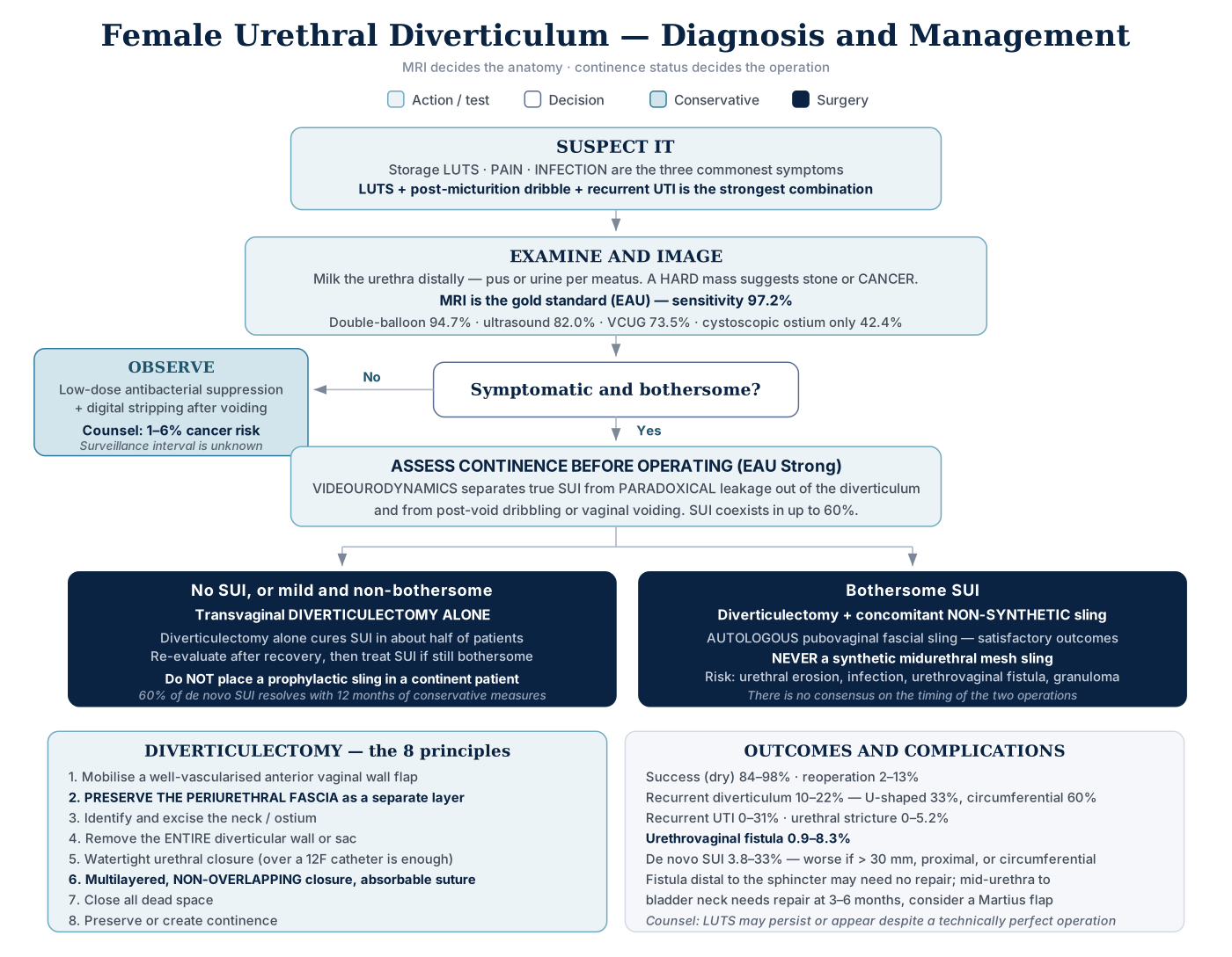
Click the algorithm to open it full size.
- A urine-filled periurethral cystic structure connected to the urethra by an OSTIUM
- Prevalence 1–6% of adult women (up to 10% in a tertiary LUTS clinic); presents in the 3rd–7th decade
- The vast majority are ACQUIRED
- From infection of the periurethral (Skene) glands → abscess → rupture into the urethral lumen → epithelialized cavity
- Up to 20% have prior urethral surgery, dilation or traumatic delivery — including after a synthetic MUS
Anatomy and histology
- Ostium is postero/ventrolateral at the 4 and 8 o'clock positions in the mid- or distal urethra in >90%
- Matching the periurethral glands
- Two-thirds of resected specimens show inflammation; neoplastic change in 6%
- Exam answer: 6–10% of diverticula harbor a malignancy; clear cell (not mucinous) is the adenocarcinoma subtype more commonly found in a diverticulum
- Female urethral adenocarcinoma likely arises from Skene's glands (female prostate homologue)
- Mucinous is commoner overall and can produce CEA
- Clear cell may secrete PSA
| Lesion | Most common malignant histology |
|---|---|
| Urethral diverticulum | ADENOCARCINOMA |
| Female urethral carcinoma | Squamous cell |
| Male urethral carcinoma | Urothelial |
Nodes
| Urethra | Proximal / posterior | Distal / anterior |
|---|---|---|
| Female | External iliac nodes | Superficial inguinal |
| Male | Pelvic nodes | Superficial inguinal |
Differential — 7 periurethral masses
- Periurethral bulking agents — also mimic a diverticulum on MRI
- Vaginal leiomyoma
- Skene gland abnormalities
- Gartner duct abnormalities
- Vaginal wall cysts
- Urethral mucosal prolapse
- Urethral caruncle
Caruncle vs prolapse: a caruncle is FOCAL, urethral prolapse is CIRCUMFERENTIAL. Caruncle = inflammatory lesion of the distal urethra in postmenopausal women → topical estrogen / anti-inflammatory cream + sitz baths; excise if large, refractory, or atypical.
Presentation
- Up to 20% are ASYMPTOMATIC
- Three commonest symptoms: storage LUTS · pain · infection
- LUTS + post-micturition dribble + recurrent UTI = the strongest combination
- Recurrent cystitis should raise the suspicion
- Others:
- Dysuria
- Hematuria
- Post-void dribbling
- Retention
- Incontinence
- Dyspareunia
- Vaginal mass
- Discharge
- Dysuria · post-void dribbling · dyspareunia = the classic "three Ds"
- Vaginal or perineal tenderness is another sign
- Vaginal pruritus is NOT a symptom
- Size does NOT correlate with symptoms — but size DOES correlate with recurrence after repair
- Mean delay to diagnosis 5.2 years across ~nine physicians, despite 52% having a palpable mass
- Examine with a speculum + careful palpation of the anterior vaginal wall 1–3 cm inside the introitus — for mass and tenderness
- Milk the urethra distally to express pus or urine. A HARD mass suggests a stone or cancer
Imaging — the sensitivity ladder
| Modality | Sensitivity |
|---|---|
| MRI — the gold standard | 97.2% |
| Double-balloon urethrography | 94.7% |
| Ultrasound | 82.0% |
| VCUG | 73.5% |
| Cystoscopy (seeing the ostium) | 42.4% |
- VCUG needs AP and lateral views
- High false-negative rate — the ostium must be patent and she must be able to void
- Double-balloon is painful, technically demanding and carries UTI risk
- Reserve for those who cannot have MRI
- Stones are found in the diverticulum 4–10% of the time
- A negative cystoscopy does NOT exclude a diverticulum
- The mid-urethral ostium (posterolateral, 4 and 8 o'clock) is seen in only 15–89% across series → proceed to T2-weighted pelvic MRI
- MRI: diverticulum is low T1 / high T2
- Independent of voiding, no radiation, and outperforms VCUG and positive-pressure urethrography
- But may miss a malignancy in the sac
Classification
- L/N/S/C3:
- Location
- Number
- Size
- Configuration
- Communication
- Continence
- Morphology:
- Simple
- SADDLEBAG (partly surrounds the urethra)
- CIRCUMFERENTIAL (completely surrounds it)
- Leng & McGuire — the surgically useful one:
- Classified by whether the periurethral fascia is preserved
- Absent/deficient fascia = PSEUDODIVERTICULUM → needs an interposition flap (Martius) or a graft such as an autologous sling
Management
- Observation: low-dose antibacterial suppression + digital stripping after voiding
- Counsel the 1–6% risk of cancer. Surveillance interval is unknown
- Intervention for symptoms: dysuria, dyspareunia, refractory post-void dribbling, recurrent UTI, pelvic pain
- 5 options:
- Excision with reconstruction (most common)
- Marsupialization (Spence-Duckett)
- Endoscopic unroofing
- Fulguration
- Incision and obliteration
- Spence-Duckett marsupialization creates a "hypospadiac" meatus
- Only for very distal lesions in women who are not sexually active
- Aggressive proximal extension risks SUI; a pseudoseptum can cause dyspareunia
Diverticulectomy — 8 principles
- Well-vascularized anterior vaginal wall flap
- PRESERVE THE PERIURETHRAL FASCIA as a separate layer
- Identify and excise the neck / ostium
- Remove the ENTIRE wall or sac
- Watertight urethral closure
- Multilayered, NON-OVERLAPPING closure, absorbable suture
- Close dead space
- Preserve or create continence
- Inverted-U incision, limbs progressively wider proximally to protect the flap's blood supply
- Inject saline, not a vasoconstrictor — so bleeding vessels declare themselves early
- Incise the periurethral fascia transversely; close the fascial flaps PERPENDICULAR to the urethral suture line
- This non-overlap is what prevents a fistula
- Urethra closes over as small as a 12F catheter without long-term stricture risk
- Three-layer closure (four with a Martius flap)
- Add a Martius flap for:
- Recurrence
- Large defect
- Deficient flaps
- Circumferential lesion needing urethral transection
- Attenuated fascia (pseudodiverticulum)
- Significant inflammation
- A planned future sling
Concomitant SUI
NEVER use synthetic mesh in an anti-incontinence procedure at the time of diverticulectomy — urethral erosion, infection, urethrovaginal fistula, granuloma.
- Bothersome SUI → concomitant NON-SYNTHETIC sling (autologous pubovaginal fascial sling) (Weak)
- VLPP 50 cm H2O (suggests ISD) → diverticulectomy + pubovaginal fascial sling, NOT a Burch colposuspension
- AUA: no synthetic midurethral sling at diverticulectomy; a Martius fat pad does NOT treat incontinence
- Do NOT place a prophylactic sling in a continent patient
- Diverticulectomy alone cures SUI in ~50% — operating on everyone is overtreatment
- Mild, non-bothersome SUI → diverticulectomy alone, then re-evaluate
- 60% of de novo SUI resolves with 12 months of conservative measures
- Question and investigate every patient for coexisting voiding dysfunction and incontinence before operating (Strong)
- Videourodynamics separates true SUI from paradoxical incontinence (urine draining out of the diverticulum) and from post-void dribbling / vaginal voiding
- There is no consensus on the timing of the two operations — diverticulectomy and any anti-incontinence procedure
- SUI coexists in up to 60%; more proximal lesions are at greater risk of post-op SUI
Outcomes and complications
- Success (dry) 84–98%, reoperation 2–13%
| Complication | Rate |
|---|---|
| Recurrent diverticulum | 10–22% — U-shaped 33%, circumferential 60% at 1 year |
| Recurrent UTI | 0–31% |
| Urinary incontinence / de novo SUI | 1.7–16.1% (de novo SUI 3.8–33%) |
| Urethrovaginal fistula | 0.9–8.3% |
| Urethral stricture | 0–5.2% |
| De novo retention | 0–9% — especially with a concomitant autologous sling |
- De novo SUI risk factors:
- >30 mm
- Proximal
- Circumferential
- Shorter urethral length
- Recurrence causes: incomplete removal · inadequate urethral closure · residual dead space in circumferential lesions
- Post-diverticulectomy urethrovaginal fistula:
- Distal to the sphincter → split stream / vaginal voiding, may need no repair
- Mid-urethra to bladder neck → incontinence → repair at 3–6 months, consider a Martius flap
- Counsel that LUTS may persist or appear despite a technically successful operation (Strong)