Neurourology
On this page
Related
Further Reading
Functional UrologyFurther Reading · Voiding DysfunctionCathetersFurther Reading · Basics- M3 receptors mediate physiologic detrusor contraction through Gq/11–phospholipase C (IP3 release of intracellular calcium) plus L-type calcium channel influx, even though M2 outnumbers M3 by at least 3:1.
- In obstructed and neurogenic bladders, the contractile subtype shifts from M3 to M2, and M3 selectivity gives little side-effect advantage because M3 also mediates dry mouth and constipation.
- Bladder relaxation during filling comes from beta-3 (and beta-2) adrenergic stimulation via the hypogastric nerve, the pathway mirabegron targets, against a background of primarily passive accommodation with parasympathetic quiescence.
- The guarding reflex is a spinal somatic reflex producing a gradual increase in striated sphincter activity (rising urethral pressure, pudendal efferent firing and periurethral EMG) during normal bladder filling.
- Glutamate is the central transmitter that stimulates micturition, driving preganglionic parasympathetic neurons from the pontine micturition center, whereas acetylcholine acts at the ganglion and the nerve–muscle junction.
- Serotonin centrally inhibits micturition, which is why duloxetine (a serotonin–norepinephrine reuptake inhibitor) helps stress urinary incontinence by boosting sacral-cord sphincter drive during filling and damping the voiding reflex.
Physiology — Storage & Emptying
Filling (Storage)
1. Accommodation
- Compliance
- Accommodation is primarily passive → intrinsic detrusor properties + parasympathetic quiescence
- Sympathetic stimulation
- β₃ (and β₂) stimulation via the hypogastric nerve → direct detrusor relaxation → the pathway mirabegron targets
- Parasympathetic inhibition
2. Closed bladder outlet
- Sympathetic stimulation → increases involuntary smooth sphincter tone
- Onuf's nucleus stimulation → increases voluntary striated sphincter tone
- Guarding reflex → spinal somatic reflex → gradual increase in striated sphincter activity during normal filling (rising urethral pressure, pudendal efferent firing, periurethral EMG)
- Serotonin centrally inhibits micturition → duloxetine (serotonin–norepinephrine reuptake inhibitor) boosts sacral-cord sphincter drive during filling + damps the voiding reflex → helps stress urinary incontinence
- Detrusor afferent input to the CNS → reflex rise in urethral pressure during filling
- Lost after orthotopic neobladder → isolated nocturnal incontinence
- Preserved after radical prostatectomy → isolated nocturnal incontinence uncommon
Emptying (Voiding)
1. Open bladder outlet
- Relaxation of the pelvic muscle
- Sympathetic inhibition
- Urethral smooth muscle relaxes → withdrawal of sympathetic excitation + parasympathetic nitric oxide release
- Onuf's nucleus inhibition
- Striated sphincter relaxes → withdrawal of pudendal tone from Onuf's nucleus
- First event in normal voiding → sudden, complete striated sphincter relaxation (falling urethral pressure, EMG silence) → seconds later detrusor contraction + vesical neck opening
- Reversal of this order = detrusor–sphincter dyssynergia
2. Detrusor contraction
- Parasympathetic stimulation
- Sympathetic inhibition
Pathways & Receptors
| System | Level | Nerve | Receptor |
|---|---|---|---|
| Sympathetic | T10–L2 | Hypogastric | β₃ |
| Parasympathetic | S2–S4 | Pelvic | M₃ |
| Somatic | S2–S4 | Pudendal (Onuf's nucleus) | — |
- Central control by the pontine micturition center
- Coordinates bladder + urethral reflex activity → suprasacral cord injury disconnects it from the sacral cord → detrusor overactivity + detrusor–external sphincter dyssynergia → upper tracts at risk
- Glutamate = central transmitter that stimulates micturition → drives preganglionic parasympathetic neurons from the pontine micturition center
- Acetylcholine acts at the ganglion + nerve–muscle junction
- Central inhibition of the micturition reflex → serotonin, plus GABA and glycine pathways at the lumbosacral cord; GABA = most widespread inhibitory transmitter in the CNS
- M₂ is the predominant receptor, but M₃ is the primary (functional) receptor
- M₂ outnumbers M₃ by at least 3:1
- M₃ drives physiologic contraction → Gq/11 → phospholipase C → IP₃ (release of intracellular calcium) + L-type calcium channel influx
- Obstructed and neurogenic bladders → contractile subtype shifts M₃ → M₂
- M₃ selectivity gives little side-effect advantage → M₃ also mediates dry mouth and constipation
Treatment Options — by Functional Category
Storage
A — Bladder related
- Behavioral → education, bladder training, timed voiding, pelvic floor
- Medication → antimuscarinic, β₃ agonist, Botox (presynaptic)
- Neuromodulation → sacral, posterior tibial
- Augmentation
B — Outlet related (sphincter)
- Behavioral therapy → bladder training, timed/fluid restriction, pelvic floor
- Sling procedure
- Artificial urinary sphincter
Voiding
A — Bladder related
- Credé maneuver
- Neuromodulation
B — Outlet related (sphincter / BPH / stricture)
- α-blocker
- Prostate surgery
- Urethral stricture repair
- Sphincterotomy
- CIC
- Indwelling catheter
EAU 16 (2025)
- EAU 16 (2025) discourages Credé / Valsalva / triggered reflex voiding:
- They raise intravesical pressure and provoke reflex sphincter contraction → hazardous to the urinary tract unless UDS confirms pressures stay within safe limits.
- All assisted emptying requires low outlet resistance + close urodynamic surveillance.
- Triggered voiding can induce AD in lesions at or above T6.
- The penile clamp is absolutely contraindicated in NDO or low compliance.
Evaluation
- Detailed history — including neurological and bowel history
- Physical exam — including sensory, reflexes, anal tone
- Voiding diary & QoL
- Labs & PVR
- Pad test & uroflow (optional)
→ After this, the patient is categorized as Low risk vs Unknown risk
Urodynamics — the only objective test of lower urinary tract function
- Video-urodynamics is the optimum invasive study in neurological patients; if unavailable → filling cystometry continuing into a pressure-flow study
- Frequency/urgency before lumbar laminectomy → cystometry first, which also sets a baseline against postoperative cauda equina compromise
- Man with hesitancy, straining, urgency and frequency after a stroke → full urodynamics before TURP or bladder neck incision, to exclude detrusor hyperactivity with impaired contractility
Risk Stratification
Framework: AUA/SUFU 2021 NLUTD. Not present in EAU 16 (2025), which uses only an informal low- vs high-risk split.
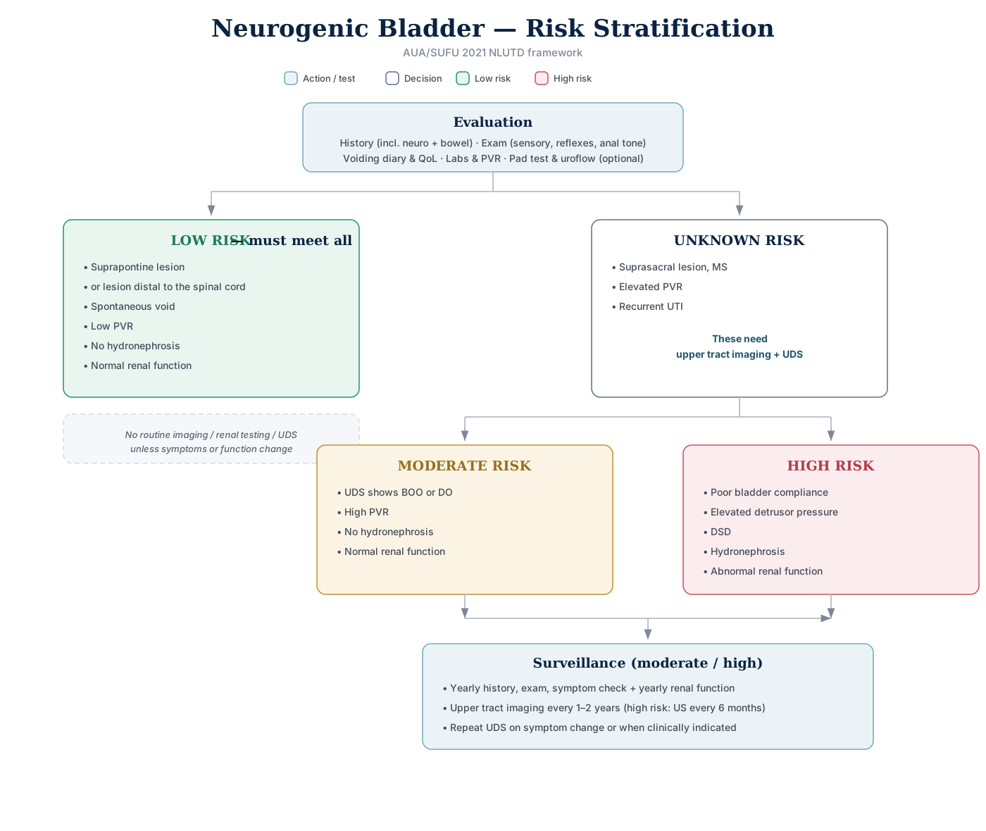
Click the algorithm to open it full size.
| Category | Defining features |
|---|---|
| Low risk (must meet all) | Suprapontine lesion or lesion distal to the spinal cord · spontaneous void · low PVR · no hydronephrosis · normal renal function |
| Unknown risk | Suprasacral lesion, MS · elevated PVR · recurrent UTI → these need upper tract imaging + UDS |
| Moderate risk | UDS shows BOO or DO · high PVR · no hydronephrosis · normal renal function |
| High risk | Poor bladder compliance · elevated detrusor pressure · DSD · hydronephrosis · abnormal renal function |
- Low risk → no routine upper tract imaging, renal function testing, or UDS
- Not clearly low risk → unknown risk until worked up, which then reclassifies as moderate or high
The four goals in a spinal cord injury patient:
- Preserve and protect the upper tracts
- Minimize lower urinary tract complications
- Treat incontinence
- Produce a management plan that is both safe and compatible with quality of life
To assess the upper tracts: a renal scan is preferred, with renal ultrasound as the alternative.
Patterns of LUT Following Neurological Disease
Key: I = impaired · OV = overactive · N = normal · S = synergic · D = dyssynergic · OP = open/incompetent · A = areflexia · CNR = competent, non-relaxing · F = fixed tone
| Disorder | Detrusor activity | Compliance | Smooth sphincter | Striated sphincter | Comment |
|---|---|---|---|---|---|
| CVA | OV | N | S | S | Suprapontine |
| Cerebral palsy | OV | N | S | S / 25% D | 25% have DSD |
| Parkinson | OV | N | S | S | Bradykinesia |
| Multiple system atrophy | OV | N | OP | S (denervation) | No TURP — open bladder neck |
| MS | OV | N | S | S / 30–65% D | 30–65% have DSD; 20–40% areflexia |
| Suprasacral cord injury | OV | N | S / D | D | Smooth sphincter DSD if lesion above T6 |
| Autonomic dysreflexia | OV | N | D | D | |
| Sacral SCI | A | N | CNR | F | Compliance may be decreased |
| Myelodysplasia / spina bifida | A | N (may be ↓) | OP | F | 10% have DSD |
| Disk | A | N | CNR | S | |
| Tabes dorsalis / pernicious anemia | I | N | S | S | Loss of sensation is the primary problem |
| Pelvic surgery | I or A | ↓ or N | OP | F | |
| Diabetes | I or A or OV | N or ↑ | S | S |
Sensation is not tabulated above but is recorded for three: CVA — may be reduced · suprasacral cord injury — absent · tabes dorsalis — loss of sensation is the primary problem.
Pattern mnemonics — which disorders cluster where:
| Finding | Mnemonic | Disorders |
|---|---|---|
| Areflexia | SIDS MD | Sacral SCI · Iatrogenic (post-op) · DM · Spina bifida · MS (20–40%) · Degenerative disk disease |
| Poor compliance | SIMS | Sacral SCI · Iatrogenic · MSA · Spina bifida |
| DSD | SMCS-T | Suprasacral SCI · MS (30–65%) · Cerebral palsy (25%) · Spina bifida (10%) · Transverse myelitis |
| Open smooth sphincter | IMS | Iatrogenic · MSA · Spina bifida |
| Competent, non-relaxing smooth sphincter | SD | Sacral SCI · Degenerative disk disease |
| Fixed external sphincter | ISS | Iatrogenic · Spina bifida · Sacral SCI |
Causes of detrusor overactivity:
- Idiopathic
- Neurogenic
- Supraspinal: stroke, Parkinson's, hydrocephalus, brain tumor, traumatic brain injury, MS
- Suprasacral spinal: SCI, cord tumor, MS, myelodysplasia, transverse myelitis
- Plus diabetes
- Non-neurogenic
- UTI
- BOO (men: BPH, bladder neck stricture; women: POP, post-incontinence surgery, primary bladder neck, stricture)
- Bladder tumor
- Bladder stones
- Foreign body
- Aging
Cerebral shock — the early period after a stroke: urinary retention from detrusor areflexia, lasting 6–12 weeks and occasionally 1–2 years. The cerebral counterpart of spinal shock.
CVA beyond the acute phase
- Detrusor overactivity with synergic relaxation of both smooth and striated sphincters and an insignificant PVR.
- External sphincter contraction during DO = a normal voluntary guarding reflex — not DESD (suprasacral SCI) and not sphincter bradykinesia (Parkinson's)
- Cerebral cortex and/or internal capsule lesions → most cannot voluntarily contract the striated sphincter, so they leak; basal ganglia or thalamic lesions spare sphincter function
- True DSD does not occur with suprapontine lesions → the EMG sphincter flare on cystometry in 5–21% of brain-disease patients is pseudodyssynergia:
- A voluntary external sphincter contraction attempting to inhibit an involuntary detrusor contraction
Cerebral palsy
- A non-progressive suprapontine brain injury
- The pontine micturition center stays intact, so the usual UDS finding is detrusor overactivity (hyperreflexia) with coordinated sphincters.
- Striated sphincter dyssynergia, when present, probably reflects additional spinal cord damage.
Brain tumor
- The superior frontal lobe is the site most often associated with voiding dysfunction
- Incontinence is the usual picture, while retention is commoner with posterior fossa tumors.
Parkinson's disease
- Mainly detrusor overactivity, but a minority have acontractility, so TURP may not help.
- Motor onset precedes LUTS by about 3 years, so the patient is already known to have PD.
- Exam answer: LUT dysfunction usually starts about 5 years after the PD diagnosis
- LUT dysfunction affects 35–70% of PD patients, with mainly storage symptoms; incontinence and large PVRs are more typical of MSA
- The DO comes with a synergic smooth sphincter; it probably reflects loss of the dopaminergic nigrostriatal pathway, which normally tonically inhibits the micturition reflex
- DO + urodynamic BPH obstruction (e.g., Pdet 88 cmH₂O, Qmax 7 mL/s, PVR 150 mL) → alpha-blocker first, before CIC or prostate surgery
- Avoid antimuscarinics until emptying improves
- Baclofen has no role — DESD is not seen in PD
- Advanced PD in retention with a low-pressure, low-flow study (detrusor underactivity) → suprapubic tube:
- CIC and neuromodulation are hard to manage, and TURP is less likely to succeed
Parkinsonism is not only PD — it is the syndrome of tremor, rigidity and bradykinesia, also produced by multiple system atrophy, progressive supranuclear palsy and Lewy body dementia.
Tethered cord
- Often declares itself at a growth spurt
- Detrusor overactivity is the commonest urodynamic finding, which is the reverse of the usual spina bifida pattern.
- Adult with spina bifida and a tethered cord → DO is still the commonest finding, even though detrusor underactivity is common in spina bifida
- Adult symptoms frequently follow activities that stretch the spine; neurosurgical therapy is controversial and may not improve the condition
Lumbar disc disease and cauda equina syndrome
- Both cause retention.
- Lumbar disc — most commonly difficulty voiding with straining and a normally compliant areflexic bladder; detrusor areflexia in 26–27% of disc protrusions
- Most protrusions compress the roots at L4–L5 or L5–S1 → sensory loss in the perineum/perianal area (S2–S4) and/or lateral foot (S1–S2); LUT dysfunction in 27–68%
- Stable L4 vertebral injury → damages only the cauda equina (the sacral cord ends at about L2) → detrusor areflexia + denervation potentials on sphincter EMG
- Because the intermingled roots also supply the external sphincter
Diabetic cystopathy
- Hits the sensory afferents first:
- Impaired sensation and increased capacity, and only later decreased contractility and a raised residual.
- Yet detrusor overactivity is the most frequent urodynamic finding.
- Early, emptying preserved (e.g., 450 mL pre-void, 60 mL residual) → timed and double voiding to prevent overdistension + intensive diabetes therapy to slow progression
Multiple System Atrophy
- Urinary symptoms — urgency, frequency and urge incontinence — can precede the diagnosis by up to 4 years.
- The bladder neck is open.
- Treating the incontinence pharmacologically may worsen emptying, so the goal is to facilitate storage and use CIC
- An indwelling catheter is considered only in advanced cases who cannot manage CIC.
- Desmopressin helps predominant nocturia.
Urogenital features favoring MSA over PD:
- Urinary symptoms, or erectile dysfunction, preceding or coinciding with the parkinsonism
- Urinary incontinence · high post-void residual · open bladder neck
- Open bladder neck at rest on videourodynamics in a man with no prior prostate surgery, especially with erectile dysfunction, is highly suggestive of MSA and separates it from PD
- Bladder neck dyssynergia instead keeps the neck closed during filling
- Striated sphincter denervation on EMG
- Worsening lower urinary tract function after urological surgery — unlike PD, where most patients do not deteriorate after surgery
TURP is an absolute contraindication in MSA (formerly Shy-Drager syndrome) — both smooth and striated sphincters are deficient, so prostatectomy removes the last outlet resistance → florid incontinence.
Autonomic Dysreflexia
- Lesion above T6–T8 — an acute, massive, disordered sympathetic response to a noxious stimulus below the level of the lesion.
- i.e., SCI at or above T6 — T6–T8 is the level of the sympathetic outflow
- It occurs only after the spinal shock phase has passed and requires a viable, intact distal cord
- The vasoconstriction is α₁-receptor mediated, and the lesion cuts descending inhibition in the lateral reticulospinal tracts.
- Mechanism — uninhibited sympathetic outflow below the lesion (splanchnic vasoconstriction) with reflex parasympathetic outflow above it.
- Hypertension = arteriolar vasoconstriction (α₁); bradycardia = an intact vagal baroreflex response to it — the vagus arises above the lesion
- Commonest trigger is bladder distension.
- Others: constipation, instrumentation, catheter change, stone, sexual activity, long-bone fracture pain, pressure sores.
- Pounding headache · hypertension · sweating and flushing above the lesion · cool skin below it · reflex bradycardia — though tachycardia or arrhythmia can occur instead.
- Urodynamics during an episode: detrusor overactivity + smooth sphincter dyssynergia + DSD.
Prophylaxis in an at-risk patient:
- Identify the at-risk patient and remove potential noxious stimuli beforehand (retention, constipation, pressure sores)
- Spinal anesthesia
- Nifedipine 10 mg, 30 minutes before the procedure — 20 mg before electroejaculation
- Labetalol · terazosin
- Routine BP + heart rate monitoring during urodynamics → early recognition; untreated AD can progress to cerebral hemorrhage or seizure
Treatment — the sequence matters:
- Remove the noxious stimulus — stop the cystoscopy, check the catheter — then sit the patient up and loosen tight clothing. These steps alone may abort the episode.
- During urodynamics: stop filling and empty the bladder first — AD from simple LUT instrumentation resolves quickly once the stimulus is withdrawn
- Drugs come only after the bladder has been emptied.
- Monitor observations every 2–5 minutes.
- Topical 2% nitroglycerin paste above the level of the lesion — standard of care.
- Alternatives: nifedipine 10–20 mg, or a beta- plus alpha-blocker.
- Persisting after bladder emptying and catheter removal:
- SBP > 150 mmHg → 2% nitropaste (½–1 inch)
- SBP < 150 mmHg → look for fecal impaction (second commonest trigger)
- Applied above the lesion because vasoconstriction below it impairs absorption; preferred as it can be wiped off if rebound hypotension occurs
- Exam answer: nifedipine is discouraged for AD treatment or prophylaxis — rebound hypotensive crises have caused stroke and MI
- Atropine 1 mg IV as needed for reflex bradycardia.
- Exam answer: atropine does not treat the hypertension — it is not the drug step when BP stays high after the bladder is emptied
- IV nitroprusside if still refractory.
- IV hydralazine is the other option for hypertension unresponsive to transdermal or oral therapy — BP becomes labile (hypotension or rebound hypertension), so admit for monitoring
For dysreflexia refractory to oral agents, ablative options exist — sympathectomy, dorsal root ganglionectomy, sacral neurectomy, sacral rhizotomy, cordectomy.
- Sacral rhizotomy with CIC is the one usually favored, being moderately invasive and relatively low risk.
EAU
- EAU defines AD as an SBP rise > 20 mmHg from baseline, with headache, blurred vision, flushing/sweating above the lesion and pallor/cold skin below it.
- It is more common in cervical (60%) than thoracic (20%) SCI; bradycardia is typical, though only 23% were actually bradycardic in one series.
- Triggers: bladder/bowel distension, cystoscopy, urodynamics, any noxious stimulus (ingrown toenail, pressure sore), sexual stimulation.
- Drugs: 2% nitroglycerin paste above the lesion is first-line; sublingual nifedipine 10–20 mg works but is prohibited in many centers (rapid, non-reversible BP drop); IV nitroprusside if refractory.
- Prophylaxis: terazosin 1–10 mg daily.
- EAU (Strong): BP + HR monitoring is mandatory during UDS and other invasive procedures in at-risk patients; empty the rectal ampulla first.
DSD — Detrusor Sphincter Dyssynergia
- Definition — a detrusor contraction concurrent with an involuntary contraction of the urethral and/or periurethral striated muscle
- On urodynamics → EMG activity during the detrusor contraction
- Pathway — neurogenic DO + DSD = disruption of the reticulospinal tract, which carries pontine micturition center output to the sacral cord to inhibit the sacral reflex and coordinate sphincter relaxation with detrusor contraction
- Suprasacral SCI
- DSD needs a lesion between the pontine micturition center (brainstem) and the sacral cord
- Suprapontine lesions typically give DO with synergic sphincters
- Sacral or more distal lesions give detrusor areflexia
- An areflexic bladder facing fixed internal sphincter activity is normal — not dyssynergia
- DSD needs a lesion between the pontine micturition center (brainstem) and the sacral cord
- MS (30–65%)
- DSD after lumbar laminectomy → suspect undiagnosed MS:
- The sacral cord begins at T12–L1, so disc herniation, cauda equina syndrome or laminectomy nerve injury are not associated with DSD
- DSD after lumbar laminectomy → suspect undiagnosed MS:
- Cerebral palsy (25%)
- Dyssynergia without a neurological lesion is not DSD → dysfunctional voiding, a learned behavior
Management
The goal is to lessen abnormal external sphincter activity or to circumvent it. Drugs used for detrusor overactivity do not relax the external sphincter — that is the central point.
- CIC
- CIC + antimuscarinics is the commonest DSD therapy. Not:
- Alpha-blockers (no effect on the striated sphincter)
- Intradetrusor onabotulinumtoxinA (lowers pressure, doesn't improve emptying)
- A male sling (raises outlet resistance)
- CIC + antimuscarinics is the commonest DSD therapy. Not:
- Sphincterotomy
- Stent across the external sphincter
- Botulinum toxin into the external sphincter
- Continuous catheterization — urethral or suprapubic
- Urinary diversion
By site of dyssynergia:
- Bladder neck → an alpha-blocker (tamsulosin)
- External sphincter → a skeletal muscle relaxant — baclofen, diazepam or dantrolene.
Indications for sphincterotomy:
- Autonomic dysreflexia caused by DSD
- DSD itself
- Risk of upper tract deterioration in a high-risk patient (DLPP > 40 cmH₂O)
- Difficulty performing CIC
- DSD with hydronephrosis/VUR, a high detrusor LPP, febrile UTIs or AD, and CIC refused or not feasible → external sphincterotomy lowers outlet resistance and LPP, allowing condom catheter drainage
- High-pressure voiding with severe hydronephrosis or VUR
- To gain the patient independence
Recurrent UTIs after sphincterotomy + condom catheter (distended bladder):
- Urodynamics first — prostatic or sphincter regrowth, stricture or lost detrusor contractility causes high-pressure voiding or incomplete emptying
- Avoid antibiotic prophylaxis, especially before bladder function is evaluated — it breeds bacterial resistance
How to decrease the rate of bladder stones in SCI with a Foley catheter
- Long-term antibiotics
- Acidify urine
- Increase fluid intake
- Routine bladder lavage
- Recurrent sediment and clogging of an indwelling catheter → urine culture first, for a urease-producing organism (e.g., Proteus mirabilis) whose biofilm drives encrustation
- Irrigation or a larger suprapubic tube only temporizes
- Urease producer found → treat it and look for bladder or upper-tract stones (cystoscopy, non-contrast CT) acting as a nidus for infection
- Recurrent sediment and clogging of an indwelling catheter → urine culture first, for a urease-producing organism (e.g., Proteus mirabilis) whose biofilm drives encrustation
Fowler Syndrome
- Urinary retention with no neurological disease in a young female
- Typically under 30, unable to void for a day or more
- Associated with PCOS
- 50% have polycystic ovaries
- Capacity over 1 L with no urge sensation
- UDS → impaired sphincter relaxation
- Abnormal external sphincter needle EMG + detrusor acontractility = diagnostic; true DSD and high-pressure, low-flow voiding are not seen
- Highly responsive to sacral neuromodulation — around 70% success, the best response of any indication
- Treatment of choice — including after failed biofeedback when CIC is declined — because it restores volitional voiding rather than bypassing the retention
- Fails: alpha-blockers (relax bladder-neck smooth muscle, not the striated external sphincter) · hormone therapy · sphincter onabotulinumtoxinA; sphincterotomy is not indicated
Impaired sphincter relaxation is the concentric-needle EMG finding (decelerating bursts / complex repetitive discharges). The urodynamic finding is detrusor acontractility.
CIC vs Indwelling Catheter
Benefits of CIC over indwelling catheter
- Improved quality of life
- Facilitates sexual activity
- Maintenance of bladder compliance — over 80% is lost with an indwelling catheter
- Upsizing the catheter, changing it more often or switching to a suprapubic tube → unlikely to improve long-term compliance
- Protect compliance / suppress DO instead → antimuscarinic or β₃ agonist, or intravesical Botox
- Avoid prophylactic antibiotics in chronically catheterized patients
- Reduced risk of pyelonephritis
- Reduced risk of stones, upper and lower tract
- Reduced risk of prostatitis, epididymitis, orchitis and Fournier's gangrene
- Reduced risk of urethral stricture
- Reduced risk of traumatic hypospadias (both sexes) and the "lead-pipe" urethra in women
UTI in SCI on CIC
- Pyuria is unreliable — catheterization alone can cause it
- Increased spasticity + bacteriuria ≥ 100 cfu/mL → symptomatic UTI → antibiotics
- Spasticity persists after treatment → look for a physical injury below the lesion (obstipation, decubitus ulcer, ingrown toenail, developing syrinx) → then baclofen
- Recurrent symptomatic UTIs → UDS for elevated storage pressures
Bladder cancer in SCI with chronic catheter
- Incidence 1% → surveillance after 8 years
Underactive Bladder (DUA)
- Difficult to diagnose — overlaps with OAB and is associated with BOO
- Diagnosis by urodynamics → measure BCI
- DUA = ↓ contractile amplitude (low Pdet + low flow) + prolonged or incomplete emptying
- High Pdet + low or normal flow → obstruction, not DUA
BCI = Pdet@Qmax + 5(Qmax)
- < 100 → underactive / acontractile bladder
- 100–150 → normal · > 150 → strong
- Unreliable when DUA and obstruction coexist (e.g. long-standing BOO)
Management
- Behavioral, timed voiding, pelvic physiotherapy
- CIC, indwelling catheter, urinary diversion
- Bladder outlet obstruction surgery
- Neuromodulation
- Bethanechol → essentially no reproducible urodynamic evidence supporting it in any patient group
- Cholinomimetic side effects: flushing, GI cramps, bronchospasm, salivation, sweating
Neuromodulation — positive results found in
- Non-obstructive urinary retention
- DUA
OAB
OAB = urgency, with or without UUI, usually with increased daytime frequency and nocturia, in the absence of UTI or other obvious pathology.
Evaluation
- Hx + physical exam + UA + PVR + QoL + voiding diary
- Urodynamics should be done if a neurological etiology is suspected
- Urodynamics & cystoscopy should be done when surgery for UUI is considered
Treatment Pathway
- Behavioral, timed voiding, fluid restriction
- Management is driven by bother → non-bothersome storage symptoms → reassurance + lifestyle changes, not drugs
- ↓ Caffeine → improves urgency and frequency, not incontinence
- 25% ↓ fluid intake → improves OAB symptoms, not UUI · warn of constipation and UTI
- Pelvic floor muscle therapy
- Urge suppression → don't rush to the toilet · sit still and contract the pelvic floor repeatedly to inhibit the detrusor until the urge passes → then walk at a normal pace
- Giggle incontinence (complete emptying on laughing, LUT function otherwise normal) → biofeedback · more durable long-term response than methylphenidate
- OAB medication (antimuscarinic / β₃ agonist)
- AUA 2nd line → start here after failed behavioral therapy (UUI, negative UA + exam) · e.g. mirabegron · UDS premature · cystoscopy not indicated
- Adequate trial → 4–8 weeks drug · 8–12 weeks behavioral → still symptomatic at 2 weeks with tolerable side effects → continue current treatment
- Failure = no improvement after 4–8 weeks or intolerable side effects → only then switch or add another oral agent
- Botox or neuromodulation
- 3rd line → SNM, Botox, PTNS · only after 2nd-line (oral) drugs fail
- Urinary diversion / augmentation
Tramadol
- ↑ Bladder capacity and compliance (animal reports) → can ease OAB symptoms
- μ-opioid effects → ↓ bladder sensation + ↑ sphincter tone → risk of postoperative urinary retention
Management of MS for LUTS
- Hx + physical exam + voiding diary + QoL + UA + US KUB + PVR + UDS
- Man with MS + DESD → renal US first — male MS patients with DESD are at greatest risk of upper tract deterioration
- Known MS + DESD → spine MRI and videourodynamics unnecessary · TRUS unneeded — the dyssynergia, not the prostate, causes the obstruction
- High PVR → CIC
- Low PVR →
- 1st behavioral therapy
- 2nd OAB medication
- 3rd Botox
- 4th neuromodulation
- 5th indwelling catheter
- 6th augmentation
MS — LUT patterns
- 5% of previously undiagnosed MS present with urinary symptoms as the first manifestation
- Voiding dysfunction → 10% at onset · 75% after 10 years
- Demyelination most often in the cervical cord — lateral corticospinal + reticulospinal columns
- NDO 43–65% (most common finding) · DSD 35% · DUA 25%
- Smooth sphincter generally synergic · striated sphincter dyssynergia coexists with DO in 30–65%
- Storage (frequency, urgency, nocturia) + voiding (hesitancy, intermittency, straining) symptoms → DO with DSD
- UDS pattern changes over time → approach irreversible treatments with caution
- Upper tract damage is rare, even with severe disability
Antimuscarinics
Divided into tertiary or quaternary.
A — Tertiary (lipophilic)
- Cross BBB → CNS side effects
- BBB crossing favored by high lipophilicity, small molecular size and low electrical charge → in a cognitively impaired patient choose a drug with low lipophilicity
- EEG → trospium no change · tolterodine occasional changes · oxybutynin consistent changes
- Hepatic metabolism → principally CYP3A4 and CYP2D6 (darifenacin, solifenacin, fesoterodine, tolterodine, oxybutynin)
- Oxybutynin (non-selective)
- Most lipophilic → may worsen cognition in elderly women
- Tolterodine
- Tissue selective (bladder > salivary gland) but no selectivity for muscarinic receptor subtypes
- Darifenacin (highest M3 selectivity)
- Relatively selective M3 antagonist, but receptor selectivity ≠ tissue selectivity (salivary glands and gut also carry M3) → dry mouth and constipation remain its main adverse effects
- Solifenacin
- Fesoterodine
B — Quaternary (lipophobic)
- Safe on CNS
- Trospium (safe, but watch the renal profile)
- Permanently charged quaternary amine → least BBB penetration → preferred in elderly cognitively impaired patients
- Trade-offs → poor GI absorption (take on an empty stomach) · dose reduction in renal impairment
- Antimuscarinic of choice in liver cirrhosis → not significantly metabolized by hepatic CYP450; eliminated mainly unchanged, actively excreted by the proximal convoluted tubules
- Also avoids interactions with CYP3A4-substrate immunosuppressants after transplantation
Contraindications
- Urinary retention
- Closed-angle glaucoma — precisely: untreated narrow-angle; treated narrow-angle is not a contraindication
- Myasthenia gravis
- cognitive impairment
Side effects
- Constipation, dry mouth, blurry vision, headache, retention
Neurogenic detrusor overactivity (NDO)
- First-line for NDO → incl. leakage between catheterizations on CIC, or poor compliance with hydronephrosis
- Ladder → antimuscarinic → botulinum toxin A → augmentation only once less-invasive treatment has failed
- Failure of one antimuscarinic does not preclude another → higher doses or combinations may be used
- Oral route poorly tolerated → intravesical oxybutynin
β₃ Agonist
- MoA → stimulates the β3-adrenoceptor → activates adenylyl cyclase → cAMP (second messenger) → detrusor relaxation during the storage phase
- Mirabegron (50 mg PO daily)
- First β3-agonist approved worldwide; little β1/β2 activity
- Increases bladder capacity without significantly changing micturition pressure, PVR or the voiding contraction
- Men with BOO → no significant difference vs placebo in Qmax or detrusor pressure at Qmax; PVR rose ~30 mL only at the unapproved 100 mg dose
- Effect at 4 weeks to 3 months
- Contraindicated → severe uncontrolled hypertension
- = systolic ≥ 180 and/or diastolic ≥ 110 mm Hg · coexisting diabetes is not a contraindication
- Measure BP before starting mirabegron and during treatment → average trial rise only ~0.5–1 mm Hg, but postmarketing reports of hypertensive crisis and cardiac / cerebrovascular events
- Vibegron → more potent and more selective than mirabegron — no measurable β1/β2 activity
- Not shown to affect BP (EMPOWUR) → urgency incontinence with memory decline + worsening hypertension → stop the antimuscarinic and use vibegron, not mirabegron
- β3-agonists have no known cognitive effect
OnabotulinumtoxinA (Botox)
- MoA → inhibits release of ACh at the presynaptic terminal
- Cleaves SNAP-25 → SNARE complex inactivated → vesicles cannot fuse → muscle paralysis (presynaptic cholinergic motor nerve endings)
- Subtype A → longest-acting of the seven botulinum toxin subtypes
- Indications → ① OAB with incontinence · ② decrease DLPP
- After injection for high DLPP → repeat UDS: safe pressures → longer catheterization intervals + reinject in 6–9 months when symptoms return · pressures still high → consider bladder augmentation
- Efficacy → 30% complete resolution · 60% have a 50% reduction in symptoms
- Retreatment often needed after 6 months
- Do not reinject within 3 months
- Complications → UTI (most common), high PVR, transient retention requiring CIC
- Retention in ~5% → worsening incontinence weeks after injection → check a PVR first to rule out overflow incontinence
- Contraindications → UTI, pregnancy, myasthenia gravis, not accepting catheterization
Steps
- Map the bladder — except the trigone, don't inject it
- 100 U / 10 mL → inject 0.5 mL × 20 sites
- Increase to 200 U / 30 mL → inject 1 mL × 30 sites — for neurogenic overactivity
- = the FDA-approved NDO dose
Sacral Neuromodulation
- MoA → modifies the voiding reflex — stimulation of the S3 afferent nerve
2-stage procedure
- Stage 1 → insertion of the electrode at the S3 root, test for 2–4 weeks; if symptoms improved →
- S3 foramen → ~9 cm from the coccyx tip, level with the inferior sacroiliac joints (11 cm is an anal-verge distance; 13 cm from the coccyx lands near S2 or S1)
- No preclinical factor (incl. urodynamics) predicts response → trial stimulation through a percutaneous or temporary lead is the best predictor of long-term response
- Stage 2 → connection of the electrode to a subcutaneous generator
How to know if placement is accurate
| Root | Response |
|---|---|
| S2 (move down) | Anus contraction + plantar flexion of the whole foot |
| S3 (target) | Bellows of the pelvic floor + dorsiflexion of the great toe |
| S4 (move up) | Bellows of the pelvic floor only — no foot or toe movement |
- S3 → bellows = levator contraction · the great toe is ipsilateral · pulling sensation in the rectum, scrotum or vagina
- Sensation → S3 and S4 both give perianal tapping · S2 gives sensation several cm from the anal canal
- S2 → also gluteal contraction, leg rotation, foot rotation, heel rotation, calf cramping, buttock sensation → move the lead one foramen lower to S3 and re-test — changing depth won't help
- S4 → move one foramen up to S3 · switch to the contralateral side only if the ipsilateral S3 response is inadequate
Complications
- Pain → most common reason for reoperation and the leading adverse event (15% at 12 months)
- Revision for an adverse event in 33% → includes relocating the generator for pocket-site pain
- Generator-site pain → turn the device off:
- Pain persists → pocket-related (infection, waistline location, pocket too tight or too loose, seroma, erosion) → needs surgery, not reprogramming
- Pain disappears → output-related → often fixed without surgery:
- Switch unipolar → bipolar programming, or reprogram around a connector fluid leak (burning)
- Revision only for intolerable symptoms
- Device infection (~3%) → pain, erythema, swelling or drainage over the generator — even if afebrile with normal labs
- Remove generator + lead (biofilm contaminates the lead) · never reimplant at the same sitting
- Reattempt only after complete healing, ≥ 6 weeks later
- Overall surgical removal → 8.5–18% of implanted patients
EAU 16 (2025)
- EAU 16 (2025) Strong: "Consider sacral neuromodulation in selected neuro-urological patients" (LE 1b) — MS with NDO are often good responders.
- MRI is no longer a barrier — current generators/leads are 1.5-T and 3-T conditional.
- SNM is not FDA-approved for neurogenic bladder (off-label) despite its efficacy — RCT success 76% on vs 42% off.
- Exam answer: SNM, while an option, is not recommended in a known neurologic condition · after onabotulinumtoxinA, consider it only once an elevated residual has been ruled out
Percutaneous Tibial Nerve Stimulation
- Insertion 5 cm cephalad to the medial malleolus and 2 cm posterior to the tibia
- Exam answer: about 3–4 cm cephalad to the medial malleolus
- Stimulates S3 through the tibial nerve
- Tibial nerve = branch of the sciatic nerve (L4–S3 fibers) → cross-talk with the sacral fibers to the bladder
- Proper placement → patient feels sensation at the bottom of the foot
- = tickling of the sole (afferent) + great-toe flexion (efferent)
- Third-line OAB therapy (2019 AUA/SUFU)
Polyuria
- Polyuria = > 40 mL/kg
- Nocturnal polyuria = 33% of the volume at night (NPI > 33%)
Evaluation
- Hx (DM, OSA, neuro) + physical + FVC (frequency-volume chart)
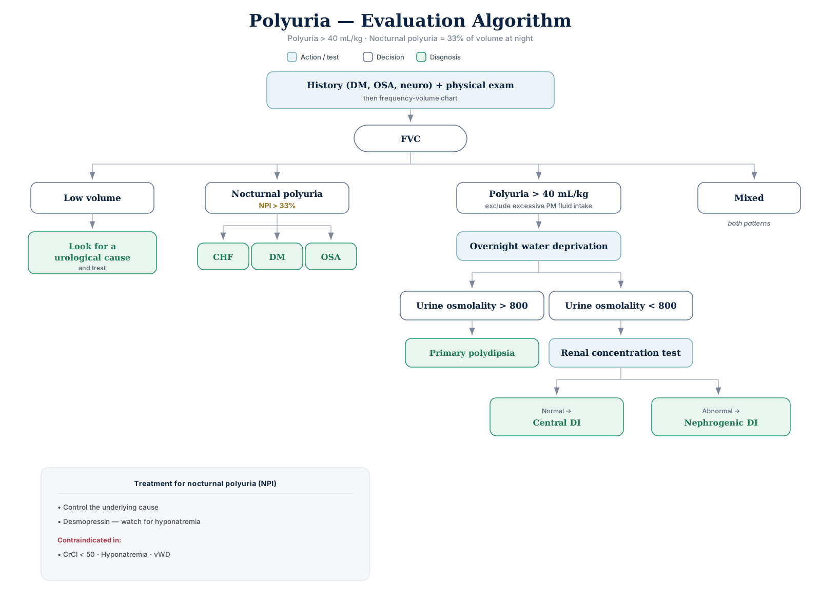
Click the algorithm to open it full size.
| FVC finding | Next step | Result |
|---|---|---|
| Low volume | Look for a urological cause | Treat |
| Nocturnal polyuria (NPI > 33%) | Look for CHF, DM, OSA, excessive PM fluid intake | Treat the cause |
| Polyuria (> 40 mL/kg) | Overnight water deprivation | Urine osmolality > 800 → primary polydipsia; Urine osmolality < 800 → renal concentration test |
| Renal concentration test | Normal → central DI | Abnormal → nephrogenic DI |
| Mixed | Both patterns |
- Global polyuria (24-h output > 40 mL/kg) with NPI < 33% → overnight water deprivation test — not alpha-blockers or antimuscarinics (neither helps global polyuria)
- Osmolality cutoff is 800 mOsm/kg; the renal concentrating capacity test is the diabetes insipidus workup
Treatment for nocturnal polyuria (NPI)
- Control the underlying cause
- Desmopressin — watch for hyponatremia
- Contraindicated in:
- CrCl < 50
- Hyponatremia
- vWD
- Contraindicated in:
Spinal Shock
- Decreased excitability of cord segments at and below the lesion → areflexic, acontractile bladder; urinary retention is the rule
- e.g. 1 week after SCI with an umbilical (T10) sensory level → large-capacity, low-pressure bladder in retention → catheterization required
- Duration → 6–12 weeks in complete suprasacral lesions; may last 1–2 years
- So at 4 weeks spinal shock still persists → UDS shows the spinal-shock picture below
- Resolution → begins with return of the bulbocavernosus reflex, alongside return of lower-limb DTRs
- First UDS within 3 months of SCI (EAU); repeat after spinal shock resolves
- Bladder management meanwhile → CIC preferred (keep storage pressure as low as achievable)
- Do not perform irreversible interventions based on symptoms during this period
Urodynamics during spinal shock:
- Detrusor areflexia; radiologically a smooth-contoured bladder without trabeculation
- Synergic smooth sphincter; closed, competent bladder neck
- Reduced but still synergic external sphincter tone — no voluntary control, and the guarding reflex is absent
- Maximum urethral closure pressure is lower than normal but maintained at the external sphincter zone
- Hypercompliance
- Absent sensation
Expected urodynamic findings by lesion:
| Lesion | Detrusor | Bladder neck | External sphincter |
|---|---|---|---|
| Complete suprasacral SCI, above T6 | Overactive | Dyssynergic | DSD |
| Complete suprasacral SCI, below T6 | Overactive | Normal | DSD |
| Complete sacral SCI | Areflexic (± poor compliance) | Tight | Fixed |
| Peripheral lesion (pelvic surgery) | Areflexic (± poor compliance) | Open | Fixed |
- Vertebral level ≠ cord segment level — the sacral cord begins at about T12–L1 and ends in the cauda equina at about the L2 vertebral level
- So a complete SCI at vertebral L2 is a sacral cord injury → competent bladder neck, but hypocontractile detrusor and no volitional external sphincter control
- Complete lesions above the sacral cord → usually DO + smooth sphincter synergy + striated sphincter dyssynergia
- e.g. T8 SCI after spinal shock → DO, smooth sphincter synergia (T8 is below the sympathetic outflow), absent guarding reflex
- Smooth sphincter dyssynergia can occur with SCI above T7 (T6 vertebral level) — the above-T6 row
Radical Pelvic Surgery
The inferior hypogastric plexus, lying alongside the rectum and vagina, is the structure at risk.
| Operation | Voiding dysfunction |
|---|---|
| Abdominoperineal resection | 20–70% · Exam answer: 20–68% (older series; lower now with nerve-sparing technique) |
| Radical hysterectomy | 16–80% |
| Low anterior resection | 20–25% |
| Proctocolectomy | 10–20% |
Injury is permanent in 15–20%.
- Presentation — a combined storage and emptying failure: inability to empty, plus stress incontinence.
- Permanent pattern = detrusor areflexia + stress incontinence — more obvious in women, as prostatic bulk masks the same urethral closure deficit in men
- Urodynamics — impaired detrusor contractility or areflexia · open smooth sphincter · fixed external sphincter (loss of voluntary control, obstructing) · reduced compliance, though it may be normal.
- Persistent pattern after APR, low anterior resection or radical hysterectomy → compliance is usually decreased (fixed striated tone keeps raising outlet resistance → poorly compliant bladder)
- The open smooth sphincter shows as an incompetent bladder neck
- Recovery takes 6–12 months. Avoid early intervention such as TURP, which can worsen the incontinence. Treatment is CIC.
- Most post-APR dysfunction is transient → no prostatectomy unless clear outlet obstruction is proven
- Retention with an acontractile detrusor after APR → continue CIC even with an enlarged prostate — TURP or Rezum risks incontinence (denervated bladder neck and membranous urethra)
- Tamsulosin and finasteride don't help — the retention is from absent detrusor contraction, not BOO
- End-fill Pdet 15 cmH₂O at 350 mL = mild compliance loss → upper tracts not at risk
- Mechanisms:
- Denervation/decentralization
- Detrusor areflexia after APR = injury to the pelvic nerves (nervi erigentes) — the preganglionic S2–S4 parasympathetic outflow running beside the rectum → bladder decentralized but not completely denervated (intramural ganglia survive)
- Tethering or encasement of nerves in scar
- Direct bladder or urethral trauma
- Bladder devascularization.
- Denervation/decentralization
Storage Pressure & the Upper Tract
- DLPP = lowest Pdet at which leakage occurs without a detrusor contraction or raised abdominal pressure
- DLPP > 40 cmH₂O → hydronephrosis or VUR in 85%
- A cut-off of 20 cmH₂O is more sensitive (91.5%) for upper tract damage
- Aim for as low a pressure as reasonably achievable, well < 40 cmH₂O — the 40 figure is not a validated "safe" line
- Impaired compliance → < 20 mL/cmH₂O (no consistent definition; 10–20 range used)
- Continent after onabotulinumtoxinA ≠ safe storage pressure → repeat UDS now; a DLPP > 40 cmH₂O reflects lost compliance and raises upper tract risk
VUR in neurogenic bladder
- Incidence 17–25% in SCI; more common with suprasacral injury
- Driven by high storage/voiding pressures + infection — no discrete VUR pressure threshold is stated
- Best initial treatment → normalize urodynamics (lower storage pressure, lower outlet resistance) before considering surgical correction
- Upper tract deterioration 51% with urethral catheters vs lower with voiding/CIC
- Chronically catheterized (e.g., tetraplegic) with new hydronephrosis → intravesical onabotulinumtoxinA (or antimuscarinics / β₃ agonists) best preserves the upper tracts → suppresses DO, maintains compliance
Risk factors for upper-tract damage in SCI:
- Bladder overdistension
- Loss of compliance
- High DLPP
- DSD.
Complications:
- VUR
- Infection (UTI, pyelonephritis)
- Stones, upper and lower
- Renal failure.
Six potentially dangerous UDS findings (usually need intervention):
- Detrusor–external sphincter dyssynergia
- Detrusor–internal sphincter dyssynergia
- Impaired compliance
- DLPP > 40 cmH₂O
- High-pressure DO throughout filling
- Poor emptying with high storage pressures.
Bladder Augmentation
- Aim → low-pressure reservoir; improves compliance, abolishes/reduces NDO
- EAU (Strong): "Offer bladder augmentation in low bladder compliance and/or refractory neurogenic detrusor overactivity" — only when all less-invasive methods have failed
- Supratrigonal cystectomy → indicated with a severely thick, fibrotic bladder wall; CIC may become necessary
- Caution with pre-operative renal scars → metabolic acidosis
- Auto-augmentation (detrusor myectomy) → low surgical burden, does not preclude further intervention
- Transplantation into a defunctionalized bladder succeeds if capacity is > 100 mL and voiding pressure < 100 cmH₂O
- Extensive pelvic irradiation → augment with transverse colon — the colon segment least likely to have been irradiated
- Irradiated bowel → anastomotic leaks, ureteroenteric strictures, poorly functioning reservoir
- Right colic artery most often arises directly from the SMA → can limit mobilizing the cecum/right colon into the pelvis; sometimes must be divided
Mitrofanoff appendicovesicostomy
- Flap valve → supple, small-caliber tube in a submucosal tunnel with firm muscular backing; rising reservoir pressure compresses it shut
- Tunnel ≥ 2 cm · channel should accept a 10–14 Fr catheter
- Flap valve grows more competent with filling; the same filling pressure tends to efface a nipple valve
- Appendiceal artery (must be preserved) → ileal branch of the ileocecal artery (an SMA branch) · anterior/posterior cecal branches → supply the cecum
Long-term complications
| Complication | Rate |
|---|---|
| Bowel dysfunction | 15% |
| Mucus production | 12.5% |
| Stone formation | 10% |
| Metabolic abnormalities | 3.35% |
| Bladder perforation | 1.9% · Exam answer: ~9% |
- Lifelong follow-up is essential — stabilizes renal function and prevents anatomical deterioration, but morbidity is significant
- Any bowel-segment reconstruction → annual history, physical, metabolic panel (electrolytes/acid-base), renal function, upper tract imaging (AUA/SUFU)
Incontinence after augmentation
- Years later, despite antimuscarinics + more frequent CIC, with negative urinalysis and normal ultrasound → videourodynamics next, not cystoscopy → assesses DO, compliance and outlet competence
- Stress incontinence on videourodynamics → outlet-enhancing procedure (e.g., sling) · DO → onabotulinumtoxinA into the native bladder or augment revision
Augment perforation
- Spontaneous perforation → most likely ischemic necrosis of the bowel segment from chronic overdistention; median ~4 years → immediate surgical repair
- Most occur in myelodysplasia → present late (impaired sensation) · risk doubles after bladder outlet procedures · recurrence up to 42%
Augment stones
- Driven by poor emptying + mucus; mostly struvite from chronic bacteriuria
- Abdominal-wall stoma → risk of incomplete emptying → best risk reduction = daily bladder irrigation + catheterization per urethra
- No proven benefit: antibiotics (may transiently lower bacteriuria, not stone risk) · thiazides · potassium citrate
Follow-up & Surveillance
No high-level evidence; intervals are largely unspecified. Flagged as such rather than invented.
EAU 16 (2025)
- Interval between baseline and control investigations should not exceed 1–2 years; much shorter in high-risk
- High risk → upper tract ultrasound every 6 months
- High risk → physical exam + urine labs yearly
- UDS → mandatory baseline in high-risk, repeated at regular intervals — no numeric interval given
- Any significant clinical change → further specialized investigation (Strong)
AUA/SUFU 2021
- Low risk → no routine yearly surveillance unless symptoms, imaging, or renal function change
- Moderate / high risk → yearly history, physical, symptom check + yearly renal function + upper tract imaging every 1–2 years
- UDS → repeat for a change of symptoms or when clinically indicated — no fixed interval
- Stone history or high stone risk → imaging every 1–2 years
Renal function (GFR) estimate
- Low muscle mass (e.g., neonates with spina bifida) → serum cystatin C is the best GFR estimate — creatinine is made only by muscle → falsely low → overestimates renal function
- Neurogenic bladder children → cystatin C is more sensitive, not more specific than creatinine
- Healthy children or non-neurogenic voiding dysfunction → normal muscle mass → similar sensitivity