Penile Cancer
On this page
- Flattening or loss of the rete pegs, caused by epidermal atrophy, is the classic histologic hallmark of lichen sclerosus (balanitis xerotica obliterans), alongside basal cell vacuolation and dermal collagen homogenization.
- Lichen sclerosus is a chronic scarring inflammatory disease that may be premalignant, linked especially to HPV-independent penile squamous cell carcinoma, and its late stage can cause penile urethral strictures.
- Penile carcinoma in situ is called erythroplasia of Queyrat on the glans or prepuce and Bowen disease on the shaft skin, genitalia or perineum, so location is the major difference.
- Unlike condyloma acuminatum, which always stays superficial regardless of size, Buschke-Löwenstein tumor invades and destroys adjacent tissue by compression, yet shows no malignant histologic change and does not metastasize.
- Kaposi sarcoma is the second most common penile malignancy after squamous cell carcinoma, because of HIV prevalence, and it also occurs with transplant or other immunosuppression.
- Lichen sclerosus can be diagnosed from its clinical appearance or confirmed by biopsy, and topical steroids are the treatment of choice for the skin disease.
Benign Penile Lesions
- Papilloma (pearly penile papules) → no treatment
- Condyloma acuminatum → HPV (6 & 11); stays superficial, never invades
- Zoon's balanitis (plasma cell balanitis) → uncircumcised men, 3rd decade+; mimics CIS → circumcision (curative)
Premalignant Lesions
| Lesion | Key | Progression to invasive |
|---|---|---|
| Giant condyloma (Buschke–Löwenstein / verrucous) | HPV 6 & 11; locally invades & destroys by compression, but does NOT metastasize | 30% |
| Bowenoid papulosis | Reddish-brown papules, shaft of young men; HPV 16 | 1% |
| CIS — Bowen disease | Penile shaft | 5% |
| CIS — Erythroplasia of Queyrat | Glans / foreskin | 30% |
| BXO (balanitis xerotica obliterans = lichen sclerosus) | — | — |
- Non-HPV also includes: penile Kaposi sarcoma (HHV-8 → screen HIV; start HAART first), leucoplakia, pseudoepitheliomatous keratotic balanitis
- Kaposi sarcoma → 2nd most common penile malignancy after SCC (HIV prevalence); also with transplant or other immunosuppression
- Kaposi in a transplant recipient → reduce immunosuppression first (often regresses) → if it fails: local excision, laser ablation or radiotherapy; no partial penectomy
- Giant condyloma vs condyloma acuminatum → condyloma acuminatum stays superficial regardless of size; giant condyloma still shows no malignant change on histology
- CIS naming = location only → glans/prepuce = erythroplasia of Queyrat; shaft skin, rest of genitalia or perineum = Bowen disease
- BXO / lichen sclerosus → chronic scarring inflammatory disease; may be premalignant (esp. HPV-independent SCC); late stage → penile urethral strictures
- Histology: flattened/lost rete pegs (epidermal atrophy) = classic hallmark; plus basal cell vacuolation, dermal collagen homogenization
- Diagnosis: clinical appearance or confirmatory biopsy; skin disease → topical steroids (treatment of choice)
- Meatal stenosis (weak stream) + non-healing glans ulcer → dilate the meatus + biopsy the ulcer (rule out malignancy)
Squamous Cell Carcinoma (SCC) of the Penis
- >95% of penile malignancies; abrupt incidence rise in 6th decade
Risk factors
- Uncircumcised
- Premalignant lesions
- HPV 16 & 18
- HPV 16 = most common genotype → 68.3% of HPV-positive tumors; next, low-risk HPV 6 (~8%)
- Chronic inflammation (lichen sclerosus, phimosis)
- Tobacco, poor hygiene/low SES, multiple partners, PUVA
Circumcision: before puberty lowers invasive cancer risk; does NOT lower risk of CIS. Adult circumcision offers little/no protection.
- Adult circumcision = after age 21 → too late, as etiologic exposure likely already occurs by puberty
- Prevention (besides prepubertal circumcision): HPV vaccination, daily genital hygiene, tobacco avoidance
Presentation
- Painless penile lesion (pain uncommon; care-seeking delay common)
- Site frequency: Glans (48%) > prepuce (21%) > shaft
- ~50% present with a palpable inguinal node (of palpable nodes, only ~43% are truly metastatic — rest inflammatory)
- Metastasis to the penis → priapism = most frequent sign; also penile swelling, nodularity, ulceration
Primary Urethral Carcinoma
- Male — site: bulbomembranous (60%) > penile (30%) > prostatic (10%)
- Commonest risk factor = urethral stricture → concentrated in the bulbar urethra
- Male — histology varies by site: anterior → up to 80% SCC; posterior → mostly urothelial; adenocarcinoma commoner in the bulbar urethra
- Female — proposed risk factors: leukoplakia, glandular metaplasia, chronic irritation, stricture, recurrent UTIs, caruncles, HPV, diverticula
- Female — histology (roughly even split): urothelial 28–45%, squamous 19–29%, adenocarcinoma 28–38%
- 6–10% of urethral diverticula harbor malignancy → adenocarcinoma predominates
- Locally advanced (T3 = invades corpora cavernosa), non-metastatic:
- SCC → curative chemoradiotherapy (genital preservation):
- 2 cycles 5-FU + mitomycin C + concurrent external beam radiotherapy → ~80% complete response, 52% 5-yr overall survival
- Urothelial → cisplatin-based chemotherapy before surgery
- SCC → curative chemoradiotherapy (genital preservation):
Staging (TNM, AJCC 8th)
T stage
| Stage | Description |
|---|---|
| Tis | Carcinoma in situ |
| Ta | Non-invasive |
| T1a | Subepithelial; no LVI/PNI and not high grade |
| T1b | Subepithelial; with LVI, PNI, or high grade (G3-4/sarcomatoid) |
| T2 | Invades corpus spongiosum |
| T3 | Invades corpus cavernosum |
| T4 | Invades adjacent structures |
- Why spongiosum = T2 but cavernosum = T3: spongiosum invasion → lower rate of inguinal node metastasis and better survival
- Urethral invasion no longer sets the T stage on its own (formerly T3) → now T2 or T3 by the corpus invaded
- A tumor near the meatus can reach the distal urethra through the spongiosum without a worse outcome
N stage
| Stage | Clinical (cN) | Pathological (pN) |
|---|---|---|
| N1 | One unilateral mobile node | ≤2 unilateral positive nodes |
| N2 | ≥2 unilateral mobile, or bilateral | ≥3 unilateral, or bilateral |
| N3 | Fixed nodal mass | Extranodal extension or pelvic node |
- Note: clinically, N3 = fixed mass; pelvic nodes are captured as pN3 pathologically — AJCC separates these. Node >4 cm often = extranodal extension.
- cN3 = fixed inguinal mass regardless of size or laterality → neoadjuvant chemotherapy before node dissection
M stage
- M1 — distant metastasis (node mets outside true pelvis, or visceral/bone).
- Most common sites: lung, bone, liver (late).
- Untreated → death in majority within 2 years.
Pathology variants
- Classic
- Verrucous → excellent prognosis (does not metastasize; observe)
- Exam answer: verrucous (Buschke–Löwenstein) rarely metastasizes → a palpable groin node is likely reactive → observe
- Biopsy only if it persists or grows
- Lymphadenectomy only for biopsy-proven metastases
- Exam answer: verrucous (Buschke–Löwenstein) rarely metastasizes → a palpable groin node is likely reactive → observe
- HPV-related
- HPV prevalence by subtype: basaloid 84% > warty-basaloid 75.7% > warty 58.7%
- p16 immunohistochemistry = surrogate marker of HPV activity → prognostic
- Sarcomatoid → very aggressive
Strongest prognostic factor for survival → extent of lymph node metastasis.
- Outranks primary tumor stage, grade and vascular invasion
- Best survival after attempted curative resection of inguinal metastases → unilateral involvement · minimal nodal disease (≤2 nodes, pN1) · no extranodal extension
Lymphatic Spread & Inguinal Anatomy
Pattern of spread
- Most common metastasis = inguinal lymph nodes (ILN):
- Penis → sentinel node → superficial nodes → deep nodes → pelvic nodes → distant mets
- Penile lymphatics can cross the midline → superficial and deep inguinal nodes → a unilateral tumor can seed contralateral nodes → bilateral inguinal nodal management is standard
- Scrotal lymphatics do not cross the median raphe → ipsilateral superficial inguinal nodes only → scrotal cancer lymphadenectomy can be ipsilateral
Inguinal anatomy (lateral → medial): "NAVEL"
| Letter | Structure |
|---|---|
| N | Nerve |
| A | Artery |
| V | Vein |
| E | Empty space |
| L | Lymphatics (→ lymph nodes) |
- Fascia lata divides superficial from deep lymph nodes
- Node of Cloquet = most cephalad deep node; marks the inguinal/pelvic boundary
ILND
- Sartorius transposition (ILND): iliac origin detached and sutured to the inguinal ligament → covers the femoral vessels and stops a superficial wound infection spreading
- Modified ILND (Catalona) → less lymphedema
- No dissection lateral to the femoral artery or caudal to the fossa ovalis · saphenous vein preserved
- Positive frozen section → mandatory conversion to standard dissection
- Number of nodes removed = main predictor of lymphatic complications → videoendoscopic and robot-assisted ILND mainly reduce wound complications, not lymphatic ones
Workup
- History & physical (incl. inguinal node exam — number, fixed vs mobile per side)
- Biopsy → assess depth, vascular invasion, grade (Broder 1–4; G1-2 = 70-80% at dx)
- Imaging →
- Small glanular lesion = none
- Larger/invasion = US (Doppler US > MRI for corporal invasion)
- CT/PET for pelvic nodes
- CT C/A/P + bone scan for distant
- Non-palpable inguinal node → DSNB (dynamic sentinel node biopsy) if ≥pT1b
- DSNB = Tc-99m nanocolloid lymphoscintigraphy + patent blue dye + intraop gamma probe
- Target false-negative rate ≤5%
- Preferred for cN0 staging.
- ~20% of cN0 groins harbor occult mets.
- Standard for detecting microscopic inguinal mets: superficial ILND or DSNB (DSNB at experienced centers)
- Sentinel node biopsy directed to a specific anatomic area (not dynamic) → unreliable for micromets, no longer recommended
- DSNB = Tc-99m nanocolloid lymphoscintigraphy + patent blue dye + intraop gamma probe
- Palpable inguinal node → FNAC/biopsy (see node algorithm)
Management of the Primary Tumor
Before choosing, identify:
- T stage: Tis/Ta · T1a/T1b · T2/T3 · T4
- Node status: non-palpable · palpable mobile <4 cm · fixed or >4 cm or N3 (= pelvis)
- Penile-sparing desired? Yes / No
- Lesion location: glans · distal · proximal
| Scenario | Treatment |
|---|---|
| Tis, Ta · N0–N2 <4 cm | Topical 5-FU, laser, wide local excision, Mohs |
| T1a, T1b · N0–N2 <4 cm · penile-sparing | Radiation or wide local excision |
| T2, T3 · N0–N2 <4 cm · penile-sparing | Radiation |
| T1–T3 · N0–N2 <4 cm · no sparing · glans | Glansectomy (if no urethral/cavernosal involvement); partial penectomy if urethra/cavernosum involved |
| T1–T3 · N0–N2 <4 cm · no sparing · distal | Partial penectomy |
| T1–T3 · N0–N2 <4 cm · no sparing · proximal | Total penectomy |
| T4, N3 or >4 cm, or M1 | Chemotherapy |
- PeIN (biopsy-proven) → first line = topical 5-FU 5% cream (or imiquimod)
- 5-FU inhibits thymidylate synthase; typically applied 12 h every 48 h × 4–6 wk
- Failed topical → don't repeat it — poor response or recurrence may signal underlying invasion
- PeIN progresses to invasive disease in 2.6–13% despite treatment (median 13 mo)
- Glans CIS, positive excision margin → treat around the scar: scar excision, laser, or topical 5-FU / imiquimod (penectomy and radiation = too aggressive)
- Foreskin SCC, pT1a (subepithelial invasion, no LVI/PNI, negative margins) → circumcision alone is definitive → surveil the glans + both groins
- Primary radiotherapy suits:
- Young men with small (2–4 cm), superficial, exophytic, noninvasive glans / coronal sulcus lesions
- Men refusing surgery
- Distant or inoperable mets needing local control
Inguinal Lymph Node Management Algorithm
Primary risk: Tis/Ta · T1a = low | T1b–T4 = high · then examine both groins (none · mobile <4 cm · fixed/>4 cm) and note laterality
POSITIVE pathway = ipsilateral radical ILND (superficial + deep) → add ipsilateral pelvic LND if ≥ 3 positive nodes on one side or extranodal extension (EAU 2026)
- AUA vs EAU: AUA adds pelvic LND at ≥ 2 positive inguinal nodes or extranodal extension
Palpable node also means a node positive on imaging (inguinal US ± FNAC, CT or PET) — treat it as cN+.
- Dissection extent: for a palpable (cN+) node, or superficial nodes +ve on frozen section in a cN0 groin → superficial + deep inguinal dissection
- Exam answer: late palpable node on surveillance (after partial penectomy, CT negative) → superficial + deep dissection on the affected side only; no antibiotics without infection concern (vs + contralateral DSNB for a unilateral node in the table below)
- Single resectable node, CT negative → dissection, not chemotherapy (no distant or unresectable disease)
Low-risk primary (Tis / Ta · T1a)
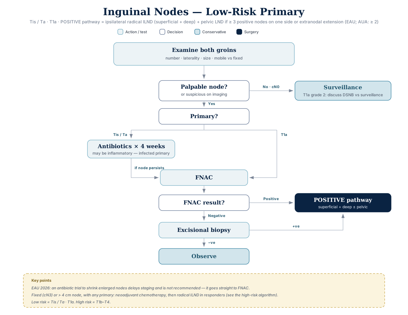
Click the algorithm to open it full size.
- pT1a G1 (grade 1 pT1, no LVI), impalpable groins → <10% inguinal met risk → observe
- Surgical nodal staging not justified — no sentinel node biopsy, no prophylactic groin radiotherapy, no lymphadenectomy
- pT1a G2 → 6–8% micrometastasis risk → surveillance can be discussed · pT1b upward → DSNB mandatory
High-risk primary (T1b–T4)
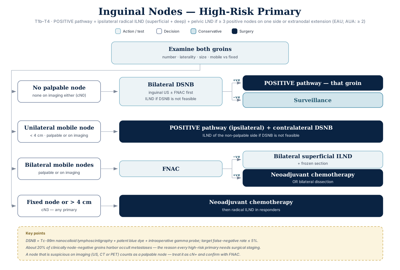
Click the algorithm to open it full size.
- T1b or higher = high risk → ~20–25% occult mets in cN0 → offer bilateral DSNB after inguinal US + FNAC; ILND only if neither DSNB nor referral is available
- Early vs delayed nodal surgery: 84% vs 35% 3-yr survival · extranodal extension 20% vs 95%
Bilateral ILND
- Exam answer: bilateral ILND is generally reserved for pT2 or high-grade tumors (vs bilateral DSNB for cN0 in the table below)
- Low-grade pT1 with LVI → consider it immediately (LVI strongly suggests occult mets)
- pT2 → bilateral ILND whether or not nodes are palpable — superficial + deep on the suspicious side, superficial on the other
- pT2: antibiotics lower the risk of inguinal wound infection, but nodal response to them should not influence the decision to stage surgically
Text version
| Primary | Node on exam | Management |
|---|---|---|
| Tis / Ta · T1a | None (cN0) | Surveillance (T1a grade 2: discuss DSNB vs surveillance) |
| Tis / Ta | Palpable | Antibiotics ×4 wk → if persists, FNAC → see FNAC outcome |
| T1a | Palpable | FNAC → see FNAC outcome |
| T1b–T4 | None (cN0) | Bilateral DSNB (inguinal US ± FNAC first; ILND if DSNB not feasible) → +ve: POSITIVE pathway on that side · −ve: surveillance |
| T1b–T4 | Unilateral mobile <4 cm | POSITIVE pathway (ipsilateral) + contralateral DSNB (ILND if DSNB not feasible) |
| T1b–T4 | Bilateral mobile | FNAC → −ve: bilateral superficial ILND + frozen · +ve: neoadjuvant chemo OR bilateral dissection |
| Any | Fixed (cN3) / >4 cm | Neoadjuvant chemo → radical ILND in responders |
- EAU 2026 differs on antibiotics: an antibiotic trial to shrink enlarged nodes delays staging and is not recommended — EAU goes straight to FNAC.
FNAC outcome (low-risk primary: Tis/Ta, T1a)
| FNAC result | Next step | Then |
|---|---|---|
| Positive | → POSITIVE pathway | — |
| Negative | → Excisional biopsy | +ve → POSITIVE pathway · −ve → OBSERVE |
- Node still palpable after 4 wk of antibiotics following excision of low-grade pT1 → FNAC (sensitivity and specificity >90%) — not observation, not immediate lymphadenectomy
- Exam answer: FNAC +ve → complete ILND on that side + superficial ILND on the opposite side
Adjuvant Chemotherapy — Indications
Consider adjuvant or neoadjuvant chemo with surgery (ilioinguinal lymphadenectomy) for:
- ≥2 positive nodes
- Exam answer: >2 positive nodes → two unilateral inguinal nodes with focal metastases is not an indication
- Bilateral metastases
- Pelvic nodes
- Extranodal extension
- Fixed inguinal masses
- Single 6-cm inguinal node
Evidence (one center): adjuvant vincristine-bleomycin-methotrexate after surgery for node-positive disease → 82% 5-year survival vs 37% with surgery alone (25 vs 31 patients)