Urethral Stricture
On this page
- Lichen sclerosus (late stage: balanitis xerotica obliterans) causes a whitish, scarred distal penis, meatal stenosis and long anterior urethral strictures, typically in men over 60.
- Lichen sclerosus is associated with penile squamous cell carcinoma, particularly HPV-negative variants, so biopsy to confirm the diagnosis and exclude malignant change, then follow up long term.
- In a woman whose urethra won't admit a 16 Fr cystoscope (normal caliber 18–30 Fr), suspect a fixed stricture from lichen sclerosus, the scarring genital-skin disease to seek on pelvic exam.
- Overall, the commonest causes of female urethral stricture are idiopathic (48.5%), iatrogenic (24.1%, mainly prior dilations, traumatic catheterization, and urethral or anti-incontinence surgery), and trauma (16.4%).
- Before meatal reconstruction for lichen sclerosus, perform retrograde urethrography, because long-standing meatal stenosis often comes with more proximal stricture disease.
- Lichen sclerosus, the commonest cause of adult meatal stenosis, is a disease of genital skin, so genital skin is unsuitable and excision with oral (buccal) mucosal graft urethroplasty is preferred.
Definitions — "stricture" is an anterior-urethra word
| Segment | Spongiosum? | Correct term |
|---|---|---|
| Anterior — penile + bulbar urethra | Surrounded by corpus spongiosum | Stricture |
| Posterior — membranous + prostatic + bladder neck | NOT surrounded by spongiosum | Stenosis / contracture; if pelvic fracture → PFUI (distraction defect) |
- Stricture = urethral epithelium + spongy erectile tissue of the corpus spongiosum → spongiofibrosis
- Anatomy that drives technique
- Bulbar urethra sits eccentrically — closer to the dorsum
- Distally the penile urethra becomes central within the spongiosum.
- (This is why a 12 o'clock DVIU is a debated incision — see the Endoluminal treatment section.)
- Corpus spongiosum has a dual blood supply
- Proximal (common penile artery, terminal branch of internal pudendal) + retrograde through the dorsal arteries as they arborize in the glans.
- (This is the whole rationale for the non-transecting rule under Urethroplasty.)
- Bulbar urethra sits eccentrically — closer to the dorsum
Etiology
Share by cause — developed-world series. Geography flips the answer: idiopathic is the commonest cause in developed countries, straddle trauma in developing ones.
| Cause | Share |
|---|---|
| Idiopathic | 33% |
| Iatrogenic | 33% |
| Post-traumatic | 19% |
| Inflammatory | 15% |
| Congenital | — |
Anterior urethra — penile & bulbar
- Straddle injury (Bulbar)
- Infection
- LS (lichen sclerosus)
- Surgery / instrumentation
- Radiation
Detail on each cause:
| Cause | Source detail |
|---|---|
| Straddle / blunt trauma | Spongiosum crushed against the inferior pubic rami. The classic mechanism of anterior stricture, and the commonest cause in developing countries |
| Iatrogenic | Cystoscopy, urethral dilation, TURP/TURBT, indwelling catheters (injury, pressure necrosis, inflammation). Very prevalent in low/middle-income countries. Silicone catheters + hydrophilic-coated ISC reduce the incidence |
| Infection | Gonococcal urethritis — historically the commonest, now rare with prompt antibiotics. Role of Chlamydia / Ureaplasma (non-specific urethritis) is unclear — no clear association established |
| LS | Strictures are longer and more often penile; higher association with urethral cancer. Involves the urethra in 20% of LS cases and is the most common cause of panurethral stricture. Also the most common cause of meatal stenosis |
| Radiation | Typically bulbomembranous. Pelvic radiotherapy strictures are usually short (mean 2.3 cm) → amenable to excision and primary anastomosis |
Lichen sclerosus in more detail
- Previously called balanitis xerotica obliterans; diagnosis is by biopsy
- Biopsy also excludes malignant change — LS is associated with penile SCC, particularly HPV-negative variants → long-term follow-up
- Appears as a whitish plaque that may involve the prepuce, glans, urethral meatus and fossa navicularis
- Late stage (balanitis xerotica obliterans): whitish, scarred distal penis + meatal stenosis + long anterior urethral stricture; typically men over 60
- If only the foreskin is involved, circumcision may be curative
- Surgery — urethroplasty with a buccal graft — is indicated in young patients with severe meatal stenosis
- LS (commonest cause of adult meatal stenosis) is a disease of genital skin → genital skin is unsuitable for reconstruction
- Excision + oral (buccal) mucosal graft urethroplasty is preferred
- LS (commonest cause of adult meatal stenosis) is a disease of genital skin → genital skin is unsuitable for reconstruction
- Because long-standing meatal stenosis often carries severe PROXIMAL stricture disease, perform a retrograde urethrogram before starting therapy
Posterior urethra — membranous & bladder neck
- Pelvic fracture
- Prostate surgery
- Radiation
- After radical prostatectomy the entity is a vesicourethral distraction defect / anastomotic stenosis
- PFUI accompanies ~10% of pelvic fractures
- the most frequent point of distraction is at the departure of the bulbous urethra from the membranous urethra
Female urethral stricture
- Fixed anatomical narrowing of the female urethra; normal caliber 18–30 Fr
- Obstructive LUTS + a 16 Fr cystoscope won't pass → suspect a fixed stricture from LS
- LS is a scarring disease of genital skin → seek it on pelvic exam (vulvar LS; also lichen planus, vulvovaginal atrophy)
Commonest causes overall — pooled series:
| Cause | Share |
|---|---|
| Idiopathic | 48.5% |
| Iatrogenic — mainly prior dilations, traumatic catheterization, urethral or anti-incontinence surgery | 24.1% |
| Trauma | 16.4% |
You should determine the following
- Length of the stricture
- Location
- Number
- density (spongiofibrosis)
How each is measured
- Length + location → radiography, urethroscopy, ultrasonography
- Depth + density → deduced from physical exam, appearance on contrast studies, and elasticity on urethroscopy.
- "The depth and density of fibrosis are difficult to determine objectively" — and the absolute length of spongiofibrosis may not be evident on ultrasound
Evaluation for stricture
- Hx + Physical Exam
- UA
- PVR (if high → Renal US & Cr)
- UFM → plateau curve
- QoL
- Urethrogram (Retrograde + Voiding)
- Cystoscopy
- cystourethroscopy reliably detects (or rules out) a stricture · also picks up lichen sclerosus and proximal bulbomembranous strictures that RUG may miss
- MRI is not beneficial for routine strictures or for pelvic fracture urethral distraction defects
- Urethral biopsy is unnecessary for a first-time urethral stricture
Two evaluation principles worth memorizing:
- "Blind passage of filiforms and blind dilation without knowledge of the anatomy of the stricture is condemned."
- In retention, if a catheter won't pass → RUG, and selectively place a suprapubic catheter to buy time for a proper plan — don't reflexively dilate.
- Evaluate the urethra completely proximal AND distal to the stricture with endoscopy + bougienage intra-operatively.
- Hydraulic pressure from voiding keeps proximal segments falsely patent; unless included in the repair they contract once the obstruction is relieved.
- in select patients, place an SPC to defunctionalize the urethra; after 6–8 weeks the hydro-dilated proximal urethra contracts and the true stricture length can be measured
- Coning down of the urethra → it is involved in the scar.
Treatment for anterior urethral stricture
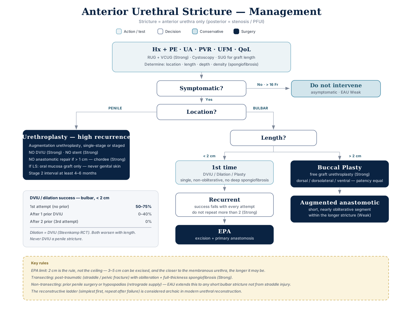
Click the algorithm to open it full size.
- Bulbar stricture management turns on LENGTH; penile stricture management turns on ETIOLOGY
- Fossa navicularis and meatal strictures are the other setting where initial endoscopic treatment is warranted — unlike the penile urethra
- Meatotomy opened to the coronal margin but lumen still 12 Fr → more than meatal stenosis
- Stop extending the urethrotomy and get a RUG to define the full stricture length (no further treatment, not even dilation, until the length is known)
- Meatotomy opened to the coronal margin but lumen still 12 Fr → more than meatal stenosis
- Should offer urethroplasty for penile stricture as high recurrence
- Do not intervene at all in asymptomatic incidental (>16 Fr) strictures — Weak [EAU]
- Consider a long-term suprapubic catheter in radiation-induced bulbomembranous strictures and/or poor performance status — Weak [EAU]
- Urethroplasty for radiation-induced stricture → success ~70% at a mean 3.5 yr follow-up · new-onset incontinence ~20% · sexual function appears unchanged
DVIU / dilation success — bulbar, <2 cm:
| Attempt | Success |
|---|---|
| 1st attempt (no prior) | 50–75% |
| After 1 prior DVIU | 0–40% |
| After 2 prior (3rd attempt) | 0% |
- Dilation = DVIU. Both get worse as length increases. Never DVIU a penile stricture.
- EPA limit: 2 cm is the rule, not the ceiling — 3–5 cm can be excised, and the closer to the membranous urethra, the longer it may be.
- Transecting: post-traumatic (straddle / pelvic fracture) with obliteration + full-thickness spongiofibrosis (Strong).
- Non-transecting: prior penile surgery or hypospadias (retrograde supply) — EAU extends this to any short bulbar stricture not from straddle injury.
- The "reconstructive ladder" (always try the simplest first, repeat after failure) is considered archaic in modern urethral reconstruction.
Endoluminal treatment — dilation & DVIU
Success rate for dilation, bulbar, for first, less than 2 cm is 50–75%. Repeated trials decrease the success. After the 3rd trial reaches 0%.
- Dilation = DVIU.
- The only high-level evidence — an RCT of n=210 with RUG at 7 visits over 48 months — showed urethral dilatation is equally effective as DVIU, and both get less effective as stricture length increases
- Goal of dilation = "stretch the scar without producing more scarring."
- If bleeding occurs, the stricture has been torn rather than stretched.
- Safest method = balloon dilating catheter over a guidewire placed under endoscopic control.
- No evidence that steroid ointment improves the outcome of urethral dilation
- DVIU technique — single incision at 12 o'clock, normal saline irrigant.
- DVIU adverse events:
- Recurrence (most common)
- Bleeding (almost always from erections immediately post-op)
- Extravasation of irrigant into perispongial tissues
- ED
- ED is the recognized complication — 5.3% (overall cold-knife complication rate 6.5%): a deep 12 o'clock incision carried beyond the spongiofibrosis can enter the corpora cavernosa
- Decreased success rate of any future reconstruction
- (deep cuts) spongiosum–cavernosum fistula
- Internal urethrotomy succeeds only when all three hold:
- The stricture is in the bulbous urethra
- It is short (<2 cm)
- It is not associated with dense, deep spongiofibrosis
- a single stricture suits DVIU best; discourage it in the penile urethra → poor patency and a higher ED risk from the incision
- Dilation may be CURATIVE in an epithelial stricture with no spongiofibrosis — the one setting where it is definitive rather than temporizing
- Catheterize for 3 to 7 days after dilation
- After uncomplicated DVIU: Foley only briefly — 3 to 5 days; EAU → remove within 72 hours · prolonged catheterization gives no advantage
- Why a catheter after DVIU at all → cover for the bleeding and irrigant extravasation listed under adverse events above
- If a short bulbar stricture recurs, a second endoscopic attempt is reasonable. Do not do a third — recurrence is effectively 100%, and it lowers the success of any later urethroplasty
- Exam answer: confirm a suspected recurrence with cystourethroscopy before any treatment
- Once confirmed → urethroplasty is preferred over repeat endoluminal treatment (see OPEN trial below)
- Exam answer: confirm a suspected recurrence with cystourethroscopy before any treatment
- Laser urethrotomy: results mixed, no better than cold knife
- Optilume (paclitaxel drug-coated balloon, FDA-approved 2023) — ROBUST III at 1 yr
- Stricture-free 74.6% vs 26.8% (p<0.0001), IPSS 9 vs 19.9, after multiple prior failed endoscopic interventions.
- Whether it works as initial rather than salvage therapy, and whether the cost is justified, is undetermined
- OPEN trial — urethroplasty vs DVIU for recurrent strictures (n=220)
- Re-intervention 13.8% vs 25.9% → 48% lower risk with urethroplasty (HR 0.52, 95% CI 0.31–0.89, p=0.017)
- 2.6× odds of a >10 mL/s Qmax gain.
- QoL similar at 2 yrs, urethroplasty costs more
Urethroplasty
Success Rate 85% even after 15 years (→ EPA). Success Rate 85% but drops to 50% after 15 years (→ Augmentation).
- With follow-up limited to 1 year, the success rate with tissue transfer clusters around 95% — but with longer follow-up it deteriorates over time.
- The success of excision and primary anastomosis at 1 year is more durable and does not deteriorate at the same rate.
EPA — for less than 2 cm, Bulbar & membranous
- "In general, 'short bulbar strictures' are those amenable to stricture excision and subsequent tension-free anastomotic repair. The limit is usually around 2 cm".
- <2 cm → EPA.
- The RCT evidence for EPA comes from strictures < 2 cm
- Best in proximal bulbar strictures → the more compliant proximal urethra can be mobilized further for a tension-free anastomosis
- Three technical points that define success — memorize as a triad:
- The area of fibrosis is totally excised
- The anastomosis is widely spatulated — a large ovoid anastomosis
- The anastomosis is tension free
- EPA reliably repairs: a PFUI with a 3 cm gap · short (1–2 cm) bulbar strictures from straddle, perineal or iatrogenic (transurethral resection) trauma
- Excision with primary anastomosis should be avoided in patients with a history of hypospadias repair
- Due to expected altered or absent retrograde blood supply to the urethra through the normal arborization in the glans
- Avoid any transecting repair here unless vessel-sparing → risk of ischemic urethral stenosis
- Use inlay or onlay grafts or flaps instead — they don't disrupt the proximal blood supply
- Vessel-sparing urethroplasty — no proven advantage over traditional EPA; reserve it for:
- Proximal supply already compromised → recurrence after a prior transecting bulbar urethroplasty
- Distal supply poor → hypospadias, especially after prior repairs
- Not the best candidate: a young man with a < 2 cm proximal bulbar stricture → standard EPA
- obliterated post-traumatic segment should be treated with transected as disturbed blood vessels will not take the graft
The bulbar ladder by length
| Length | Repair |
|---|---|
| < 2 cm | End-to-end anastomosis — mobilize both ends · remove the scar · spatulate · evert · anastomose over a catheter · close the spongiosum |
| 2–4 cm | Anastomotic repair is still possible, but in older patients tissue elasticity is less — use a buccal graft |
| > 4 cm | Substitution urethroplasty |
- Exam answer: a 3 cm bulbar stricture → free graft (buccal mucosa) substitution urethroplasty
- Even in a young man — especially if distal bulbar, where the urethra is less compliant
- EPA would create tension and ischemia at the anastomosis
- Dilation / DVIU / laser urethrotomy → little to no success
- Graft placement in substitution repair depends on where the stricture sits:
- Proximal bulbar → graft VENTRAL
- Distal bulbar → graft DORSAL
Augmentation — General Rule
Graft is better than flap
- Dorsal buccal onlay beats a fasciocutaneous flap → fewer complications · better patient satisfaction · better long-term success
- Dorsal onlay is better for diverticulum issue
- Single-stage over staged repair when feasible → similar long-term outcomes with fewer operations
- e.g. a recurrent 3 cm bulbar stricture in a urethroplasty candidate → single-stage dorsal onlay buccal mucosa graft
- DVIU is not first-line > 2 cm (dilation + self-catheterization only for non-candidates)
- e.g. a recurrent 3 cm bulbar stricture in a urethroplasty candidate → single-stage dorsal onlay buccal mucosa graft
- Use buccal or lingual mucosa if a graft is needed and available
- Use penile skin if buccal/lingual is not available, suitable, or accepted
- Do not use genital skin graft in case of LS
- Do not use hair-bearing perineal or scrotal flaps unless no other option
- Four grafts used for primary urethral reconstruction:
- Oral mucosal graft (buccal / labial / lingual)
- Buccal mucosa is thought to have a panlaminar plexus — this is why it takes so well.
- Bladder epithelial graft
- Rectal mucosal graft
- Full-thickness skin graft. (STSG is for staged repair.)
- Oral mucosal graft (buccal / labial / lingual)
What makes buccal mucosa the graft of choice
- Easy accessibility · hairless · resistant to infection
- Thick epithelium — promotes easy handling
- Thin, well-vascularized lamina propria — promotes inosculation
- Compatible with a moist environment
Graft has 3 phases to take
- Imbibition — 48 hrs
- Hours 0–48 · the graft "drinks" nutrients from the host bed · graft temperature below core body temperature
- Inosculation — 48 hrs — microcirculation
- Hours 48–96 · microcirculation restored · graft rises to core body temperature → at 72 h the graft is inosculating
- Revascularization
- Exam answer: take lasts about 96 h in two phases (imbibition → inosculation); revascularization is the process of take itself
- Take depends on both the nature of the grafted tissue and the host bed → anything that impairs host-bed vascularity impairs take
- Take needs immobilization and close contact with a well-vascularized bed → why grafts are quilted
- Classic causes of graft failure:
- Seroma
- Hematoma
- Infection
- Scar tissue
- Classic causes of graft failure:
- Graft vs flap — the definition:
- A flap is transferred WITH its blood supply (preserved or surgically re-established)
- A graft is transferred to a host bed where a new blood supply develops by take.
- i.e. tissue excised from a donor site that reestablishes its blood supply by revascularization — that process is take
Skin grafts for penile shaft coverage
- After debridement for Fournier's gangrene → resurface the shaft with an unmeshed, thick (0.012–0.015 inch) split-thickness skin graft (STSG) → highest take and best cosmesis
- Meshed or thinner STSG → contracts more → compromises penile function and cosmesis
- Full-thickness graft → fails to take more often
- Abdominal or thigh flaps → poorer cosmesis
Ways to gain more length — especially in EPA transecting in pelvic fracture case
- Mobilize to the level of the suspensory ligament
- Separate the crura of the corpora
- = corporal splitting — the first move when the ends still won't meet tension-free after mobilization · gains length without removing bone
- Split in the midline over 4–6 cm, up to where the corpora fuse · bleeding here = the deep dorsal vein of the penis → suture-ligate it
- Inferior pubectomy
- Only after corporal splitting → risks penile shortening · destabilized erection · pelvic destabilization with chronic exercise-related pain
- Route the urethra around corpora
- Supracrural rerouting — the last resort, needed in < 10% of uncomplicated cases
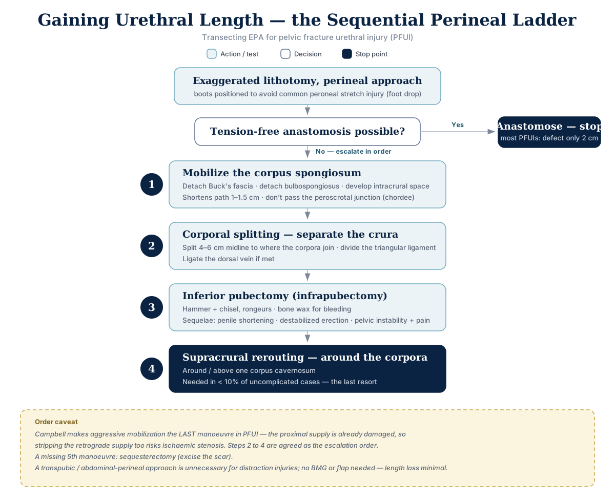
Click the algorithm to open it full size.
Order caveat: step 1 is standard, limited mobilization; aggressive mobilization is best kept as the LAST maneuver in PFUI — the proximal supply is already damaged, so stripping the retrograde supply too risks ischemic stenosis. Steps 2 to 4 are agreed as the escalation order. A missing 5th maneuver: sequesterectomy (excise the scar). A transpubic or abdominal-perineal approach is unnecessary for distraction injuries; no BMG or flap is needed — length loss is minimal in most cases.
- These steps (spongiosal mobilization → crural separation → inferior pubectomy → rerouting) = the progressive perineal approach → closes even longer posterior gaps tension-free
- So a combined transpubic abdominoperineal approach is usually not needed
Deep dorsal vein of the penis — the vein between the corpora
- Lies beneath Buck's fascia, between the paired dorsal arteries · main drainage of the glans and the distal two-thirds of the corpora cavernosa
- Runs behind the symphysis pubis → drains into the periprostatic (Santorini) plexus
Posterior urethra / PFUI — management
Timing
- Deferred repair at earliest 3 months after trauma — by then the pelvic hematoma has resolved, the prostate has descended into a more normal position, the scar has stabilized, and the patient can lie in lithotomy.
- "If possible, it is desirable to proceed within 3 to 6 months, after trauma"
Endoluminal treatment of posterior stenosis
| Recommendation | Strength |
|---|---|
| Do not perform endoscopic treatment for an obliterative stenosis | Strong |
| Perform one attempt at endoluminal treatment for a short, non-obliterative stenosis | Weak |
| Do not perform >2 DVIU/dilatations for a short non-obliterative recurrence after EPA for traumatic posterior stenosis, if long-term patency is the intent | Weak |
Rationale: "cut-to-the-light" on an obliterative stenosis will not work and risks a false passage towards the bladder base or rectum.
- Anastomotic recurrence after posterior urethroplasty:
- Non-obliterative and <1 cm → DVIU
- After two failed DVIUs/dilatations → redo urethroplasty (three or more incisions are never successful)
- Incising a membranous recurrence:
- Avoid deep 6 o'clock cuts (rectal injury) and deep 12 o'clock cuts (urosymphyseal fistula)
- Warn of de novo incontinence in up to 25% (worsening of existing incontinence in up to 15%)
Technique & outcomes
- Repair: most PFUIs are short → mobilization of the corpus spongiosum + primary anastomotic technique via a perineal approach (λ-shaped incision, exaggerated lithotomy).
- Anastomosis of the proximal anterior urethra can be made to any segment of the posterior urethra — apical, prostatic, or below
- Technique of choice for a PFUI on suprapubic diversion (posterior urethra almost always obliterated) → deferred (≥3 months), one-stage perineal excision and primary anastomosis (EPA)
- ~10% of PFUIs are complex and may have fistulae — most commonly urethrorectal
- Post-op:
- Urine diverted via the SPC; the small silicone urethral catheter is plugged and serves as a stent only.
- Bed rest 24–48 h.
- Voiding trial with contrast at 3–4 weeks
- Void urethrally for 5–7 days, then remove the SPC.
- Flexible endoscopy at ~6 months and again at 1 year
- Curative rates for posterior PFUI reconstruction: high 90% range
- Exam answer: perineal EPA for obliterative posterior stenosis → overall stricture-free rate 85.7%
- Aligning catheters:
- Total circumferential distraction often does not occur — a strip of epithelium is often left intact, and an aligning catheter may let it heal virtually unscarred.
- An aligning catheter is a guide, not a traction device.
- It also drains the liquefying pelvic hematoma.
- But many still stricture after removal and need definitive repair
- Some recommend against routine use because realignment is associated with delayed surgical repair and more unsuccessful endoscopic treatments
Penile urethral strictures
| Recommendation | Strength |
|---|---|
| Do not use DVIU for penile strictures | Strong |
| Do not use urethral stents for penile strictures | Strong |
| Offer men with penile urethral stricture disease augmentation urethroplasty by either a single-stage or staged approach, considering previous interventions and stricture characteristics | Strong |
| Do not offer anastomotic urethroplasty to patients with penile strictures >1 cm — due to the risk of post-operative penile chordee | Strong |
| Counsel patients with penile strictures that single-stage procedures might be converted to staged ones in the face of adverse intra-operative findings | Strong |
| Offer an interval of at least 4–6 months before the second stage, provided the first stage outcome is satisfactory | Weak |
- The one to carry away: EPA is contraindicated in penile strictures >1 cm (chordee).
- Why: the pendulous urethra has no redundancy → excision shortens it relative to the corpora → ventral chordee
- First-line for a penile stricture = buccal graft augmentation urethroplasty — not dilation or urethrotomy (high recurrence)
- Contrast bulbar and fossa/meatal strictures, where initial endoscopic treatment is warranted
One-stage vs two-stage
- One stage — when the cause is trauma, instrumentation or catheter-related.
- The choice depends on surgeon experience, tissue status, circumference and the site of the stricture.
| Approach | Technique | When |
|---|---|---|
| Flap | Orandi | Mid-shaft stricture with a free external urethral meatus |
| Flap | Jordan | Meatus involved |
| Graft (buccal) | ASOPA | Any site or length, meatus involved or not |
- Two stage — when the cause is failed or crippled hypospadias, lichen sclerosus, or radiotherapy.
- Stage one opens the urethral plate, removes all fibrous scar and lays the graft
- Stage two tubularizes the plate around the catheter.
- Stage it when the urethral plate is inadequate for a single-stage repair, or with heavy scarring, LS, fistula or chordee; staged (mainly buccal) repairs report 71–95% patency
- Failed hypospadias repair with scarred penile skin → two-stage buccal mucosal graft urethroplasty (oral mucosa = graft of choice)
- The interval before stage two lets the graft heal before tubularization
The onlay hierarchy
- Onlay procedures — graft or flap — have a higher success rate than tubularized grafts or tubularized skin islands
- Among tubularized repairs, tubularized flaps do better than tubularized grafts
- Dorsal and lateral onlay are likely better than ventral onlay
- In the penile urethra a full-thickness skin graft beats a split-thickness graft — STSG contracts and causes chordee
- Most common complications of penile urethroplasty: fistula and meatal stenosis
Perineal urethrostomy (PU)
The escape hatch for complex disease — failed hypospadias repairs, LS, multiple failed reconstructions, elderly/comorbid patients.
| Recommendation | Strength |
|---|---|
| Offer PU as a management option to men with complex anterior urethral stricture disease | Strong |
| Offer PU for men not fit or not willing to undergo formal reconstruction | Weak |
| Choose the type of PU based on personal experience and patient characteristics | Weak |
| Consider augmented Gil-Vernet-Blandy PU or "7-flap" PU in men with proximal bulbar or membranous stricture disease | Weak |
- Why not just reconstruct?
- Pananterior reconstruction with multiple grafts is feasible with fair success — but success rates fall with multiple failed urethroplasties, and complex prolonged surgery may not be indicated in the elderly/comorbid
- Long penile stricture after multiple failed repairs → PU ~90% success vs ~80% for two-stage urethroplasty; single-stage repair is inappropriate without healthy penile urethral tissue (success = long-term patency, no CIC, good patient-reported outcomes)
- Permanent PU for long-segment strictures when urethroplasty is unsuitable (the complex-disease patients above) or self-dilation is impossible — e.g., Parkinsonian tremor
- Complex reconstruction adds little over PU but more perioperative risk
- BMG-augmented PU — for stricture continuing into the proximal bulbar and/or membranous urethra, where traditional PU can't work.
- Also indicated after pelvic radiation or LS, which have a higher rate of re-stricture at the PU.
- The graft is spread-fixed to the corpora (same bed as a dorsal onlay) and decreases the probability of circumferential scarring at the neomeatus.
Other urethral pathology
- Urethral hemangioma
- Electrofulguration only to control an acute episode
- Smaller lesions — laser, which produces less scarring: argon, KTP (532 nm), or Nd:YAG
- Larger lesions — open excision and urethral reconstruction, sometimes circumferential
- Avoid tubed GRAFT reconstruction. A tubed flap or mixed tissue transfer may be considered, though staged reconstruction is probably preferable
- Polidocanol as a sclerosing agent has good initial success in extensive lesions
- Reactive arthritis — the classic triad of arthritis, conjunctivitis and urethritis.
- Urethral involvement is usually mild and self-limited, and a minor part of the disease.
- Urethral amyloidosis — rare
- Consider it in any patient with a urethral mass.
- Presents with hematuria, dysuria or obstruction.
- Urethrocutaneous fistula — an epithelium-lined tract from urethra to skin.
- Follows urethral surgery, or develops secondary to periurethral infection with an inflammatory stricture, or after treatment of a urethral growth.
- Treatment must address the underlying process, not just the defect.
- Congenital urethral diverticulum — a transitional-cell-lined pouch
- Arising from distension of a segment or from attachment by a narrow neck.
- In males a "congenital" anterior diverticulum may reflect incomplete urethral development, or straddle trauma causing an intracorporeal spongiosal hematoma
- A congenital diverticulum in the PROSTATIC urethra is a remnant of the müllerian duct
Hematuria — localizing the source
- Initial hematuria → most often the prostatic and anterior urethra (blood sitting in the urethra is flushed out by the first part of the stream)
- Terminal hematuria → trigone, bladder neck or prostate (bleeds as the bladder neck contracts at the end of voiding)
- Total hematuria → bladder or upper tract (blood mixes throughout the stored urine)
- Clot shape: vermiform clots → upper tract source; cuboid clots → bladder source
- Hematuria that clears after catheter placement → more likely prostatic in origin
Complications of urethroplasty
| Complication | Rate / detail |
|---|---|
| Permanent ejaculatory dysfunction | As high as 20% after urethroplasty. Semen pools in the urethra and/or loss of force with ejaculation. |
| Temporary ED | Up to 20% after anterior urethroplasty — same for EPA and graft/flap repairs. Resolves classically by 6 months, with <3–4% permanent. Overall rate of ED after urethroplasty ≈ rate after circumcision. Longer-segment reconstructions → higher risk, though function improves over time. |
| Recovery of potency | Directly related to preoperative potency — the repair itself rarely causes lasting ED |
| New-onset penile curvature | Usually after an over-aggressive EPA in the distal bulbar region |
| Post-void dribbling | Bulbospongiosus midline division may damage muscle + perineal nerves. A muscle- and nerve-sparing perineal approach has been introduced to reduce it |
| In PFUI | ED in PFUI is often a consequence of the injury itself, not the surgery |
| ED treatment after trauma | Wait 2 years before definitive ED treatment such as a penile prosthesis — injury-related ED can still recover in that time |
| SUI after PFUI urethroplasty | More likely with post-trauma de novo ED — the same nerve injury (prostate torn from the bulbar urethra) impairs the involuntary bladder neck sphincter. Age, prostate size and defect length are not associated. Open bladder neck on the preoperative filling cystogram → bladder neck sphincter dysfunction and SUI risk; closed bladder neck → intact sphincter |