Urodynamics (UDS)
On this page
Related
Further Reading
Functional UrologyFurther Reading · Voiding DysfunctionFlowcharts
Female Stress Urinary IncontinenceClinical flowchart · Voiding DysfunctionOveractive BladderClinical flowchart · Voiding DysfunctionNon-neurogenic Male LUTSClinical flowchart · Voiding DysfunctionUnderactive BladderClinical flowchart · Voiding DysfunctionNeuro-urology (NLUTD)Clinical flowchart · Voiding DysfunctionFemale Bladder Outlet ObstructionClinical flowchart · Voiding Dysfunction- The five reference uroflow curve types are bell shaped, tower, plateau, staccato, and intermittent, per the International Children's Continence Society 2014 terminology standardization.
- Poorly compliant bladders, including those with bladder outlet obstruction, show an increased ratio of type III to type I collagen and a raised connective tissue to smooth muscle ratio in the bladder wall.
- Once collagen has replaced the bladder stroma, reduced compliance usually does not respond to drugs, hydraulic distension or nerve section, and augmentation cystoplasty is often needed.
- Historically, detrusor overactivity (then called detrusor instability) required an involuntary detrusor pressure rise greater than 15 cm H2O during filling at a bladder volume of 300 mL or less.
- The current ICS definition of detrusor overactivity is any involuntary detrusor contraction during filling cystometry, with no pressure threshold; 40 cm H2O instead marks a dangerous detrusor leak point or sustained storage pressure.
- Intermittent large-volume leakage with normal compliance, a low PVR and no stress leak suggests detrusor overactivity, which urodynamics fail to show in about 50% of patients with urgency incontinence.
What the Study Is
| Tier | Components |
|---|---|
| Standard | Uroflowmetry + PVR + cystometry + pressure-flow study |
| Supplementary | EMG + imaging (videourodynamics) with continuous urethral pressure |
Including: UFM · PVR · cystometry · PFS · EMG · cystogram
- Pdet = Pves − Pabd
- Pves = bladder catheter
- Pabd = rectal (or vaginal/stomal) catheter — use rectal when possible; data are limited for vaginal and stomal
- Subtraction is what separates a true Pdet rise (contraction or lost compliance) from a Pabd artifact (straining, Valsalva, movement)
Indications for UDS
- Helpful while diagnosis remains uncertain
- Mixed symptoms, or who failed previous treatment
- Urinary retention or high PVR
- Prior to any invasive procedure
EAU framing of the same idea
| Recommendation | Strength |
|---|---|
| Adhere to ICS good urodynamic practice standards | Strong |
| Do not routinely perform UDS for uncomplicated SUI | Strong |
| Do not routinely perform UDS before first-line treatment of uncomplicated OAB | Strong |
| Perform UDS if findings may change the choice of invasive treatment, and/or if there is diagnostic uncertainty | Weak |
PFS Is Indicated Before Invasive Treatment
- Previous unsuccessful invasive treatment for LUTS
- Patient can't void more than 150 mL
- 80 years or older with voiding symptoms
- Younger than 50 years with voiding symptoms
- Qmax > 10 mL/s when considering surgery
- with Qmax < 10 mL/s, BOO is likely and UDS is not necessarily needed — the test earns its place when flow is equivocally preserved
- PVR > 300 mL when considering invasive therapy
The 9 Cs — Parameters to Record
Filling cystometry
- Coarse sensation
- Capacity
- Compliance
- Contraction (involuntary detrusor)
- Continence
Voiding PFS
- Contractility
- Clinical obstruction
- Complete emptying
- Coordination
Uroflowmetry (UFM)
| Parameter | Value |
|---|---|
| Voided volume (VV) — minimum volume | 150 mL |
| Qmax — likely obstruction | < 10 mL/s |
| Qmax — suspect obstruction | 10–15 mL/s |
- Qmax < 10 mL/s for BOO: specificity 70%, PPV 70%, sensitivity 47% — rules in, doesn't rule out
- Qmax has within-subject variation — repeat the study, especially if VV < 150 mL or the pattern is abnormal
- Instruct the patient not to Valsalva void
- Voiding efficiency = VV / (VV + PVR) × 100
- Do not offer non-invasive tests as an alternative to UDS/PFS for diagnosing BOO in men
Pattern of UFM
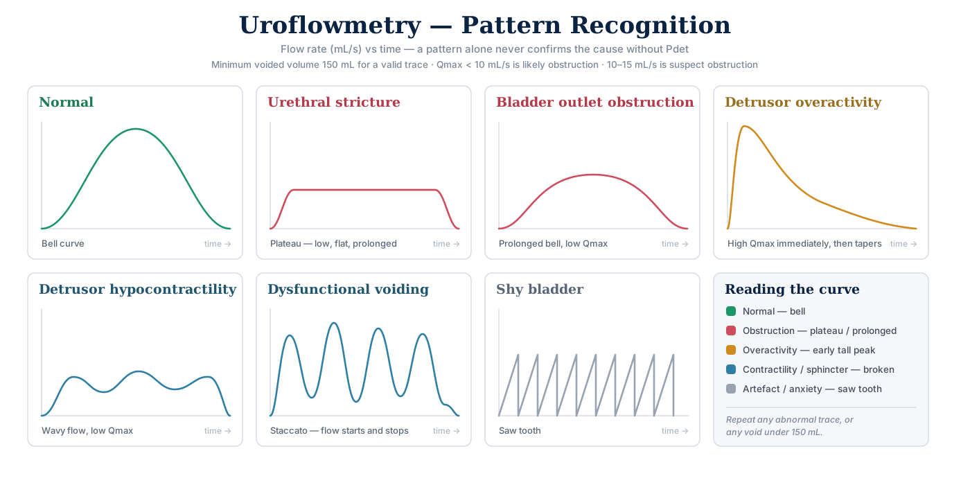
Click the chart to open it full size.
- The pattern suggests, it never confirms
- Curve interpretation is subjective; confirm a staccato/intermittent trace on non-invasive uroflow to exclude a test-induced phenomenon
Pattern nomenclature
| Pattern | Suggests |
|---|---|
| Bell | Normal |
| Flattened | Fixed obstruction — the plateau trace above |
| Interrupted / straining / wavy | Impaired contractility, obstruction, or abdominal straining |
| Peaked / tower | Rapid emptying, overactivity — closest to the high Qmax then tapers trace |
| Staccato | Dysfunctional voiding — "intermittent or fluctuating flow rate secondary to involuntary intermittent contractions of the periurethral striated muscle in neurologically normal individuals" |
| Saw tooth | "Shy bladder" |
- Five reference curve types (International Children's Continence Society, 2014 terminology standardization):
- Bell-shaped
- Tower
- Plateau
- Staccato
- Intermittent
Cystometry (Filling)
| Parameter | Value |
|---|---|
| Filling rate | (wt/4) ≈ 20–30 mL/min |
| Capacity | ≈ 400–500 mL normal adult |
| Compliance | = Δ Volume / Δ Pressure — normal 46–124 mL/cm H₂O; impaired if < 20 |
| Detrusor pressure | (contraction) |
| Continence | LPP |
- Filling rate: physiologic = filling rate below the predicted maximum of body weight (kg) / 4 mL/min; above that is non-physiologic.
- For a 80–120 kg adult, wt/4 lands exactly on your 20–30 mL/min.
- After pelvic radiation: reduced bladder capacity is the most common urodynamic finding
- Sometimes with reduced compliance and detrusor overactivity — radiation effects evolve and may progress over time
Compliance — the practical rules
- Absolute pressure beats the compliance number.
- "In practical terms, absolute pressure is probably more useful than a 'compliance number'" — storage pressure > 40 cm H₂O harms the upper tracts
- Why: a 400 mL bladder at Pdet 50 and a 40 mL bladder with an early IDC both compute to ~6–8 mL/cm H₂O — only the first is dangerous. The number alone cannot tell them apart
- ICS measures two points: Pdet at start of filling (usually zero) and Pdet at cystometric capacity — both excluding detrusor contractions
- So in practice compliance = volume / end-fill Pdet → e.g. 300 mL at 6 cm H₂O = 50 mL/cm H₂O → normal
- Causes of impaired compliance (4):
- Neurologic (SCI, spina bifida — usually via increased outlet resistance/DESD or decentralization)
- Long-term BOO
- Radiation cystitis
- TB
- Wall change: poorly compliant bladders (including BOO) show ↑ type III : type I collagen and ↑ connective tissue : smooth muscle ratio
- Once collagen has replaced the stroma, reduced compliance usually does not respond to drugs, hydraulic distension or nerve section → augmentation cystoplasty is often needed
- False positive (looks worse than it is): rapid filling → stop filling; if pressure returns to baseline, compliance is not impaired
- False negative (looks better than it is): "pop-off" mechanisms — VUR and bladder diverticula.
- With VUR the pressure is transferred to the refluxing kidney and is harmful; VUDS is very useful here
Detrusor overactivity (DO)
- Definition: involuntary detrusor contraction during the filling phase. Any phasic contraction during filling is DO regardless of amplitude
- This is the current ICS definition → 40 cm H₂O is not a DO cutoff; it marks a dangerous detrusor leak point or sustained storage pressure
- Historically ("detrusor instability"): required an involuntary Pdet rise > 15 cm H₂O during filling at a volume ≤ 300 mL
- Neurogenic (relevant neurological condition) vs idiopathic · spontaneous vs provoked · single vs multiple · phasic / sporadic / terminal
- Terminal DO = a single involuntary contraction at cystometric capacity causing incontinence
- Non-phasic Pdet change before micturition = a compliance change, not DO
- Failure to demonstrate DO does not rule it out — detection is influenced by patient position
- UDS miss DO in ~50% of patients with urgency incontinence
- Clue on a negative study: intermittent large-volume leakage + normal compliance + low PVR + no stress leak → suggests DO
- Be certain the contraction is truly involuntary — patients get confused and void on first desire
- Prognostic: OAB with obstruction is more likely to resolve after TURP when DO is a single terminal IDC rather than continuous/sporadic
Leak Point Pressures
Continence, LPP
- DLPP → to assess risk of upper tract deterioration, if more than 40 cm is at risk
- ALPP → to assess SUI, if less than 60 it is due to ISD, if more than 100 it is not due to ISD
- Exam answer: ALPP measures sphincter strength — < 60 cm H₂O suggests ISD · 60–90 equivocal · > 90 little or no ISD
| ALPP | DLPP | |
|---|---|---|
| Measures | Sphincter response to increased Pabd | Injured bladder response to increased outlet resistance |
| Definition | Pves at which leakage occurs, from raised Pabd, without a detrusor contraction | Lowest Pdet at which leakage occurs, without raised Pabd or a detrusor contraction |
| Direction | Lower = weaker sphincter | Higher = more dangerous to upper tracts |
| Question answered | Is this SUI due to ISD? | Are the upper tracts at risk? |
ALPP
- There is no normal ALPP — patients without SUI will not leak at any physiologic Pabd. Only demonstrable in a patient with SUI
- Measure the total Pabd required to cause leakage, not the change — includes baseline Pabd (usually 20–40 cm H₂O standing).
- Read from the Pves channel provided there is no involuntary contraction
- Start at 150 mL, then every 50 mL until SUI is demonstrated. If none at capacity → remove the urethral catheter and measure via the abdominal channel
- Sphincteric SUI testing needs volume ≥ 200 mL and Valsalva Pabd ≥ 60 cm H₂O — missing either is a main cause of a false-negative study
- Post-prostatectomy SUI missed despite adequate volume and Valsalva
- → the catheter can occlude the bladder neck or cause sphincter spasm: remove it at capacity and repeat Valsalva
- Grade 3 cystocele → falsely high ALPP — it kinks and obstructs the urethra.
- Reduce it during UDS (pessary, swab or split speculum) → unmasks occult SUI and gives the true ALPP
- ALPP ≠ VLPP. In the same person, Valsalva LPP < cough LPP
- Current technology cannot distinguish ISD from urethral hypermobility in women.
- If there is no hypermobility, SUI must be ISD regardless of ALPP — an isolated ALPP has limited utility in predicting surgical success
AUA vs EAU — the role of LPP. AUA/Campbell use ALPP to characterize ISD and to inform choice of anti-incontinence surgery ("clinicians making the diagnosis of urodynamic SUI should assess urethral function" — ALPP or MUCP). EAU 14: "Do not use urethral pressure profilometry or leak point pressure to grade severity of urinary incontinence" — Strong
DLPP
- The significance: bladder pressure is climbing too high before the pop-off of urethral leakage occurs.
- Low outlet resistance → leak at a safe pressure
- High outlet resistance (e.g. DESD) → pressure keeps climbing and reaches the kidneys — less incontinence, more danger
- Most useful in (3):
- UMN lesions with high storage pressures (DO + DESD)
- LMN disease causing decentralization
- Non-neurogenic low compliance (multiple bladder surgeries, radiation, tuberculous cystitis)
- After sphincterotomy: DLPP is the most reliable urodynamic predictor of upper tract deterioration — > 40 cm H₂O = the sphincterotomy has failed → guides whether to repeat it
- Urethral function tests (urethral pressure profile, Valsalva LPP, EMG) show no correlation with upper tract deterioration
- Abnormal compliance on CMG is worrisome, but a much less established predictor
Voiding PFS
Records: Qmax, curve, PVR
| Parameter | Value |
|---|---|
| Pdet | 40–60 normal, female less |
| EMG | ? DSD — look for |
| BOOI | = Pdet@Qmax − 2(Qmax) → ≥ 40 means obstructed (full bands below) |
| BCI | = Pdet@Qmax + 5(Qmax) → < 100 means weak detrusor contractility (full bands below). Weak is not acontractile — an acontractile detrusor shows no demonstrable contraction at all on UDS, a distinct ICS term |
The three fundamental patterns
| Pdet | Flow | Diagnosis |
|---|---|---|
| Low / normal | High / normal | Normal, unobstructed voiding |
| High | Low / normal | Obstruction |
| Low | Low | Detrusor underactivity |
Full bands
| Index | Value | Meaning |
|---|---|---|
| BOOI | ≥ 40 | Obstructed |
| 20–40 | Equivocal | |
| ≤ 20 | Unobstructed | |
| BCI | > 150 | Strong contractility |
| 100–150 | Normal contractility | |
| < 100 | Weak contractility |
BOOI/BCI caveats
- Both indices break down when underactivity and obstruction coexist — e.g. long-standing obstruction
- BOOI grossly underestimates female BOO (females void at much lower pressures)
- → female cutoffs: Qmax ≤ 12 mL/s + Pdet@Qmax ≥ 25 cm H₂O — but with large overlap; needs radiographic or clinical corroboration
- BOO also damages storage: ~⅔ of men with symptomatic BOO have DO, resolving 50–67% of the time with treatment of the obstruction. Reduced compliance also improves after TURP
- Significantly impaired compliance remains the only absolute urodynamic indication for treating BOO — a critical BOOI level has never been defined
- If there is no obstruction on PFS, surgical outcomes are less favorable
Orthotopic neobladder voiding
- Voids by Valsalva with external sphincter relaxation — the bowel reservoir has no coordinated detrusor contraction
- Pattern: Pabd ↑ · Pves ↑ · Purethra ↓ — straining raises abdominal pressure, transmitted to the pouch, while urethral pressure falls
- Emptying can fail over time: flaccid bowel wall or an abdominal-wall hernia → can't raise pouch pressure enough → some need intermittent catheterization
Coordination — EMG & Sphincter
- Normal: EMG activity decreases before a voluntary bladder contraction. Failure to relax, or to stay relaxed, during micturition is abnormal
- EMG lag time: normal 2–6 s with a bell-shaped voiding curve · < 2 s common in OAB · > 6 s → primary bladder neck dysfunction
- Pseudodyssynergia: EMG sphincter "flare" during filling from voluntarily trying to inhibit an involuntary contraction (guarding reflex) — not true dyssynergia
- DESD: involuntary increase in external sphincter activity with detrusor activity/voiding
- True DESD requires a known neurologic lesion between the pons and the sacral micturition center (infrapontine, suprasacral — Onuf's nucleus S2–S4)
- No neurologic lesion → it is learned behavior = dysfunctional voiding, not DESD
- Detrusor contracting against a closed sphincter → high pressures → can impair compliance → urodynamic risk factor for upper tract deterioration
- DISD: lack of coordination between detrusor and internal sphincter/bladder neck
- Dysfunctional voiding (the learned, non-DESD pattern above) = external sphincter spasm during voiding
- Pelvic floor muscle dysfunction (perineal pain, constipation, pelvic muscle tenderness)
- → most common UDS finding is a low peak flow with an intermittent pattern — outlet obstruction from the sphincter spasm
- DO, poor compliance and large bladder capacity are not typical — unless chronic over several years
- Neurologically intact woman with a staccato (intermittent) uroflow, incomplete emptying and recurrent UTIs
- → increased EMG activity during voiding confirms non-neurogenic voiding dysfunction or Fowler's syndrome
- Pelvic floor muscle therapy (teaches pelvic relaxation) is often first-line for either; several studies show sacral neuromodulation is effective
- Pelvic floor muscle dysfunction (perineal pain, constipation, pelvic muscle tenderness)
Dangerous UDS Findings — Usually Require Intervention
Six findings that threaten upper and lower tract decompensation:
- Impaired compliance
- Detrusor–external sphincter dyssynergia (DESD)
- Detrusor–internal sphincter dyssynergia (DISD)
- Detrusor leak point pressure > 40 cm H₂O
- High-pressure detrusor overactivity present throughout filling
- Poor emptying with high storage pressures
These are the findings that justify treating on the tracing alone, independent of symptoms — impaired compliance from radiation cystitis or TB can only be diagnosed by CMG
Videourodynamics
- Procedure of choice for confirming bladder neck dysfunction in men and women
- Primary bladder neck obstruction can only be diagnosed on VUDS
- Differentiates functional obstruction (PBNO vs dysfunctional voiding) — AUA/SUFU supports VUDS in young men and women without an obvious anatomic cause of obstruction
- Especially useful for BOO in women; can confirm sphincteric dysfunction diagnosed by EMG
- Man with perineal discomfort, Qmax 9 mL/s and PVR 100 mL despite antibiotics and an alpha-blocker
- → VUDS to diagnose bladder neck obstruction
- Synergistic voiding on uroflow EMG rules out pelvic floor dysfunction
- In presumed bladder neck obstruction, amitriptyline worsens voiding and pelvic floor physiotherapy is not beneficial
- Young man with severe LUTS, Qmax 8 mL/s and PVR 95 mL despite tamsulosin
- → VUDS before any surgery — low flow fits either obstruction (including PBNO) or detrusor underactivity
- Other uses (5):
- Localize obstruction
- Detect incontinence not seen on exam
- Evaluate VUR during storage/voiding
- Determine whether a known anatomic abnormality (diverticulum, VUR) is contributing
- Neuropathic voiding dysfunction
- Refer to a VUDS center if a complete diagnosis can't otherwise be obtained:
- Known/suspected neurogenic LUTD
- Unexplained female retention
- Prior radical pelvic surgery
- Urinary diversion
- Pre/post renal transplant
- Prior pelvic radiation
- Ambulatory UDS: more physiologic, best when standard UDS is inconclusive and treatment is uncertain
Conducting the Study — Practical
- Decide on/off medication in advance:
- Testing the drug's effect → study on a regular dosing schedule.
- Uncovering the cause of symptoms after empirical treatment → consider stopping first for the highest yield
- UDS is an unnatural setting:
- Failure to record an abnormality does not rule it out
- Not all UDS observations are clinically significant
- Ideally the patient's symptoms are reproduced during the study
- Sedation / anesthesia: DO is the only parameter reliably affected — may be absent on a sedated study
- Compliance, detrusor leak point pressure and bladder capacity are generally unaffected
- Define the question before you start. If it isn't answered, repeat the study in the same session — many authors advocate a second filling and/or voiding cycle
- Antibiotic prophylaxis is recommended for UDS in patients with indwelling urethral/suprapubic catheters, condom catheters, or on CIC
- Air-charged vs water-filled catheters:
- Air-charged avoid hydrostatic effects and motion artifact and need no leveling at the symphysis
- But nomograms were built on fluid-filled systems and readings are systematically higher — not interchangeable